Page 555 - Atlas of Small Animal CT and MRI
P. 555

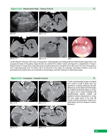
Figure 5.4.8  Adenomatous Polyp—Pylorus (Canine)                                            CT


            (a) US, SP                       (b) CT+C, TP


            (c) CT+C, TP                     (d) CT+C, TP                     (e) ES
            14y MC Miniature Schnauzer with history of regurgitation. Ultrasonography was initially performed, which showed a hyperechoic mass
            occupying the lumen of the pylorus. Images b and c are ordered from caudal to cranial, and image d is a magnification of image c.
            Delayed‐contrast CT images show a hypoattenuating to  isoattenuating mass in the pyloric antrum (b: arrows). The mass is heterogeneous
            with a hyperattenuating center. The pyloric wall is not thickened in this region. An endoscopic examination revealed a mural mass
              protruding into the pyloric lumen (e: arrowheads). Excisional biopsy confirmed a diagnosis of adenomatous polyp.


              Figure 5.4.9  Leiomyoma—Stomach (Canine)                                                    CT

                                                                             17y MC mixed‐breed dog. Images are ordered
                                                                             from cranial to caudal. A gastric mass was
                                                                             identified on screening ultrasound. The well‐
                                                                             defined mass on the medial wall of the  fundus
                                                                             has multifocal mineralized regions (a: arrow).
                                                                             The gastric wall is focally thickened with
                                                                             hypoattenuating contents caudally and
                                                                             peripheral contrast enhancement (c: arrow).
                                                                             The most caudal aspect of the mass (d)
                                                                             remains centrally unenhanced. Surgical exci-
                                                                             sional biopsy confirmed a diagnosis of gastric
            (a) CT+C, TP                     (b) CT+C, TP
                                                                             leiomyoma.


            (c) CT+C, TP                     (d) CT+C, TP
                                                                                                             545
   550   551   552   553   554   555   556   557   558   559   560