
Page 553 - Atlas of Small Animal CT and MRI
P. 553
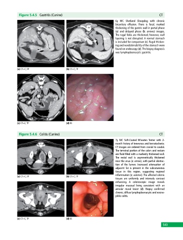
Figure 5.4.5  Gastritis (Canine)                                                            CT

                                                                             6y MC Shetland Sheepdog with chronic
                                                                             bicavitary effusion.  There is focal, marked
                                                                             thickening of the gastric wall in portal phase
                                                                             (a) and delayed phase (b: arrows) images.
                                                                             The rugal folds are thickened; however, wall
                                                                             layering is not disrupted. A normal stomach
                                                                             is included for comparison (c). Rugal thicken-
                                                                             ing and nondistensibility of the stomach were
                                                                             found on endoscopy (d). The biopsy diagnosis
                                                                             was lymphoplasmacytic gastritis.


            (a) CT+C, TP                     (b) CT+C, TP


            (c) CT+C, TP                     (d) ES


              Figure 5.4.6  Colitis (Canine)                                                              CT

                                                                             7y MC Soft‐Coated  Wheaten  Terrier with 2‐
                                                                             month history of tenesmus and hematochezia.
                                                                             CT images are ordered from cranial to caudal.
                                                                             The terminal portion of the colon and rectum
                                                                             are fluid filled with a markedly thickened wall.
                                                                             The rectal wall is asymmetrically thickened
                                                                             near the anus (c: arrow), with partial obstruc-
                                                                             tion of the lumen. Increased attenuation of
                                                                             adjacent fat is present in the subcutaneous
                                                                               tissue in this  region,   suggesting regional
                                                                             inflammation (c: asterisk). The affected colonic
            (a) CT+C, TP                     (b) CT+C, TP
                                                                             tissues are uniformly and intensely contrast
                                                                             enhancing.  A colonoscopic image reveals
                                                                               irregular mucosal lining consistent with an
                                                                             annular mural lesion (d). Biopsy confirmed
                                                                             chronic, diffuse lymphoplasmacytic and eosino-
                                                                             philic colitis.


            (c) CT+C, TP                     (d) ES
                                                                                                             543