
Page 551 - Atlas of Small Animal CT and MRI
P. 551
Gastrointestinal Tract 541
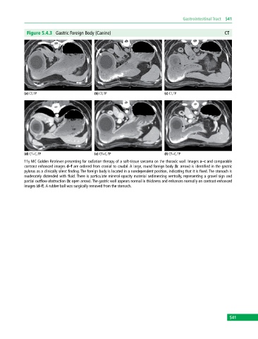
Figure 5.4.3 Gastric Foreign Body (Canine) CT
(a) CT, TP (b) CT, TP (c) CT, TP
(d) CT+C, TP (e) CT+C, TP (f) CT+C, TP
11y MC Golden Retriever presenting for radiation therapy of a soft‐tissue sarcoma on the thoracic wall. Images a–c and comparable
contrast enhanced images d–f are ordered from cranial to caudal. A large, round foreign body (b: arrow) is identified in the gastric
pylorus as a clinically silent finding. The foreign body is located in a nondependent position, indicating that it is fixed. The stomach is
moderately distended with fluid. There is particulate mineral‐opacity material sedimenting ventrally, representing a gravel sign and
partial outflow obstruction (b: open arrow). The gastric wall appears normal in thickness and enhances normally on contrast‐enhanced
images (d–f). A rubber ball was surgically removed from the stomach.
541