
Page 544 - Atlas of Small Animal CT and MRI
P. 544
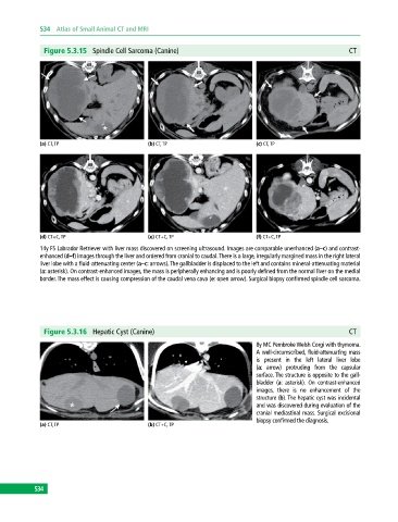
534  Atlas of Small Animal CT and MRI


              Figure 5.3.15  Spindle Cell Sarcoma (Canine)                                                 CT


             (a) CT, TP                       (b) CT, TP                       (c) CT, TP


             (d) CT+C, TP                     (e) CT+C, TP                     (f) CT+C, TP
             14y FS Labrador Retriever with liver mass discovered on screening ultrasound. Images are comparable unenhanced (a–c) and contrast‐
             enhanced (d–f) images through the liver and ordered from cranial to caudal. There is a large, irregularly margined mass in the right lateral
             liver lobe with a fluid‐attenuating center (a–c: arrows). The gallbladder is displaced to the left and contains mineral‐attenuating material
             (a: asterisk). On contrast-enhanced images, the mass is peripherally enhancing and is poorly defined from the normal liver on the medial
             border. The mass effect is causing compression of the caudal vena cava (e: open arrow). Surgical biopsy confirmed spindle cell sarcoma.


              Figure 5.3.16  Hepatic Cyst (Canine)                                                         CT
                                                                               8y MC Pembroke Welsh Corgi with thymoma.
                                                                               A well‐circumscribed, fluid‐attenuating mass
                                                                               is present in the left lateral liver lobe
                                                                               (a: arrow) protruding from the capsular
                                                                                 surface. The structure is opposite to the gall-
                                                                               bladder  (a:  asterisk).  On  contrast‐enhanced
                                                                               images, there is no enhancement of the
                                                                                 structure (b). The hepatic cyst was incidental
                                                                               and was discovered during evaluation of the
                                                                                 cranial mediastinal mass. Surgical excisional
                                                                               biopsy confirmed the diagnosis.
             (a) CT, TP                       (b) CT+C, TP


           534