
Page 542 - Atlas of Small Animal CT and MRI
P. 542
532 Atlas of Small Animal CT and MRI
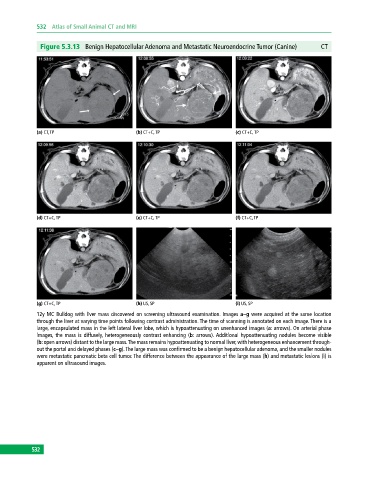
Figure 5.3.13 Benign Hepatocellular Adenoma and Metastatic Neuroendocrine Tumor (Canine) CT
(a) CT, TP (b) CT+C, TP (c) CT+C, TP
(d) CT+C, TP (e) CT+C, TP (f) CT+C, TP
(g) CT+C, TP (h) US, SP (i) US, SP
12y MC Bulldog with liver mass discovered on screening ultrasound examination. Images a–g were acquired at the same location
through the liver at varying time points following contrast administration. The time of scanning is annotated on each image. There is a
large, encapsulated mass in the left lateral liver lobe, which is hypoattenuating on unenhanced images (a: arrows). On arterial phase
images, the mass is diffusely, heterogeneously contrast enhancing (b: arrows). Additional hypoattenuating nodules become visible
(b: open arrows) distant to the large mass. The mass remains hypoattenuating to normal liver, with heterogeneous enhancement through-
out the portal and delayed phases (c–g). The large mass was confirmed to be a benign hepatocellular adenoma, and the smaller nodules
were metastatic pancreatic beta cell tumor. The difference between the appearance of the large mass (h) and metastatic lesions (i) is
apparent on ultrasound images.
532