
Page 539 - Atlas of Small Animal CT and MRI
P. 539
Hepatobiliary Disorders 529
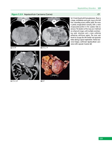
Figure 5.3.9 Hepatocellular Carcinoma (Canine) CT
9y FS Dachshund with hemoabdomen. There is
a large, multilobular and cystic mass in the left
liver, extending across midline (a,b). The mass
is poorly encapsulated cranially, with a more
encapsulated portion in the caudate lobe (c:
arrows). The borders of the mass are ill defined
on enhanced images, with multiple unenhanc-
ing cystic structures and a more uniformly
enhancing soft‐tissue region. The mass was
found to occupy the left medial and quadrate
lobes during surgical exploration. Partial exci-
sional biopsy confirmed hepatocellular carci-
noma with capsular invasion (d).
(a) CT, TP (b) CT+C, TP
(c) CT+C, DP (d) GP
529