
Page 537 - Atlas of Small Animal CT and MRI
P. 537
Hepatobiliary Disorders 527
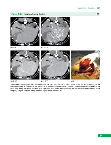
Figure 5.3.6 Hepatic Adenoma (Canine) CT
(a) CT, TP (b) CT+C, TP
(c) CT+C, TP (d) CT+C, TP (e) GP
10y FS mixed‐breed dog with elevated liver enzymes. The liver mass is located in the left lateral lobe and is hypoattenuating on the
unenhanced image (a: arrowheads). The gallbladder (a: asterisk) is displaced by the mass effect. There is contrast enhancement of the
entire mass during the arterial phase (b), mild hyperattenuation on the portal phase (c), and isoattenuation on the delayed phase
image (d). Surgical excisional biopsy confirmed hepatocellular adenoma (e).
527