
Page 528 - Atlas of Small Animal CT and MRI
P. 528
518 Atlas of Small Animal CT and MRI
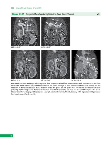
Figure 5.2.15 Congenital Extrahepatic Right Gastric–Caval Shunt (Canine) MR
(a) T1+C, 3D, DP (b) T1+C, 3D, DP
(c) T1+C, 3D, DP (d) T1+C, 3D, DP (e) T1+C, MIP, DP
4mo M Yorkshire Terrier with suspected portosystemic shunt. Images are ordered from ventral to dorsal (a–d) after subtraction. The shunt
arises at the cranial aspect of the gastroduodenal vein (b: GD), travels from right to left in the cranial abdomen (a–d: arrows), and then
terminates in the caudal vena cava (d: C). The shunt crosses the splenic and left gastric veins but does not anastamose with them
(a: S,LGV). The MIP image shows the course of the shunt in its entirety (e: arrows). See page 507 for Legend for Figures 5.2.1–5.2.18.
Clinic of Small Animal Surgery and Reproduction, Ludwig Maximillian Universität, Munich, Germany, 2014. Reproduced with permission
from Ludwig Maximillian Universität.
518