Page 527 - Atlas of Small Animal CT and MRI
P. 527

Hepatovascular Disorders  517


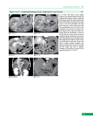
              Figure 5.2.14  Congenital Extrahepatic Shunt—Right Gastric–Caval (Canine)                   CT
                                                                              1y FS  Terrier with failure to gain weight.
                                                                              Images a–c are ordered caudal to cranial, and
                                                                              images d–f are ordered cranial to caudal. The
                                                                              white arrows denote the cranial course of the
                                                                              shunt and the black arrows denote the caudal
                                                                              course to the shunt termination. The large
                                                                              shunt vessel (a–c: white arrow) arises from
                                                                              the gastroduodenal vein at the level of the
                                                                              right gastric vein and travels cranially to the
                                                                              porta hepatis, where it gives off a right intra-
                                                                              hepatic branch (b: arrowhead). It crosses to
            (a) CT+C, TP                     (b) CT+C, TP
                                                                              the left side (a–c: black arrow) and travels
                                                                              caudally to join the caudal vena cava near the
                                                                              right kidney (d: black arrow). The dorsal plane
                                                                              MIP image shows the right (e: white arrows)
                                                                              and left (e: black arrows) arms of the shunt
                                                                              vessel.  A  plastic  ameroid  constrictor  was
                                                                              placed around the shunt vessel at its junction
                                                                              with the  caudal  vena  cava  (f:  asterisk),
                                                                              occluding the blood flow. See page 507 for
                                                                              Legend for Figures 5.2.1–5.2.18.


            (c) CT+C, TP                     (d) CT+C, TP


            (e) CT+C, MIP, DP                (f) CT+C, TP


                                                                                                             517
   522   523   524   525   526   527   528   529   530   531   532