
Page 522 - Atlas of Small Animal CT and MRI
P. 522
512 Atlas of Small Animal CT and MRI
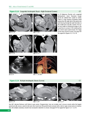
Figure 5.2.8 Congenital Intrahepatic Shunt—Right Divisional (Canine) CT
1y FS Doberman Pinscher with suspected
portosystemic shunt. Transverse images
(a–d) are ordered from caudal to cranial.
There is a large, tortuous anomalous vessel
arising from the right side of the portal vein
and traveling through the right liver lobes to
the caudal vena cava (b,c: arrows). The ves-
sel was visible on ultrasound images in the
cranial liver (e: arrows). A 3D rendering from
the ventral aspect demonstrates the shape
of the shunt vessel (f: arrow). See page 507
(a) CT+C, TP (b) CT+C, TP
for Legend for Figures 5.2.1–5.2.18.
(c) CT+C, TP (d) CT+C, TP
(e) US, OP (f) CT+C, 3D, VENT
Figure 5.2.9 Multiple Intrahepatic Shunts (Canine) CT
(a) CT+C, TP (b) CT+C, TP (c) CT+C, TP
6mo MC Labrador Retriever with failure to gain weight. Preoperatively, there are multiple small, tortuous vessels within the hepatic
parenchyma (b: arrows), cranial to the main shunt vessel (a). After partial shunt occlusion with a stent (c: open arrow) and coil emboliza-
tion, the multiple anomalous vessels have increased in diameter (c: arrows). See page 507 for Legend for Figures 5.2.1–5.2.18.
512