
Page 519 - Atlas of Small Animal CT and MRI
P. 519
Hepatovascular Disorders 509
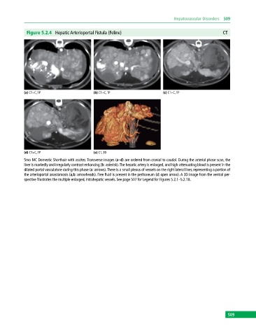
Figure 5.2.4 Hepatic Arterioportal Fistula (Feline) CT
(a) CT+C, TP (b) CT+C, TP (c) CT+C, TP
(d) CT+C, TP (e) CT, 3D
5mo MC Domestic Shorthair with ascites. Transverse images (a–d) are ordered from cranial to caudal. During the arterial phase scan, the
liver is markedly and irregularly contrast enhancing (b: asterisk). The hepatic artery is enlarged, and high‐attenuating blood is present in the
dilated portal vasculature during this phase (a: arrows). There is a small plexus of vessels on the right lateral liver, representing a portion of
the arterioportal anastomosis (a,b: arrowheads). Free fluid is present in the peritoneum (d: open arrow). A 3D image from the ventral per-
spective illustrates the multiple enlarged, intrahepatic vessels. See page 507 for Legend for Figures 5.2.1–5.2.18.
509