
Page 509 - Atlas of Small Animal CT and MRI
P. 509
Body Wall, Retroperitoneum, and Peritoneal Cavity 499
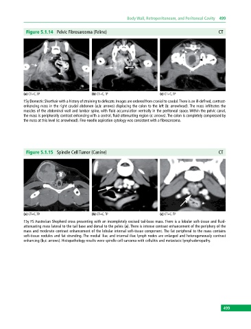
Figure 5.1.14 Pelvic Fibrosarcoma (Feline) CT
(a) CT+C, TP (b) CT+C, TP (c) CT+C, TP
15y Domestic Shorthair with a history of straining to defecate. Images are ordered from cranial to caudal. There is an ill‐defined, contrast‐
enhancing mass in the right caudal abdomen (a,b: arrows) displacing the colon to the left (b: arrowhead). The mass infiltrates the
muscles of the abdominal wall and lumbar spine, with fluid accumulation ventrally in the peritoneal space. Within the pelvic canal,
the mass is peripherally contrast enhancing with a central, fluid‐attenuating region (c: arrows). The colon is completely compressed by
the mass at this level (c: arrowhead). Fine‐needle aspiration cytology was consistent with a fibrosarcoma.
Figure 5.1.15 Spindle Cell Tumor (Canine) CT
(a) CT+C, TP (b) CT+C, TP (c) CT+C, TP
15y FS Australian Shepherd cross presenting with an incompletely excised tail‐base mass. There is a lobular soft‐tissue and fluid‐
attenuating mass lateral to the tail base and dorsal to the pelvis (a). There is intense contrast enhancement of the periphery of the
mass and moderate contrast enhancement of the lobular internal soft‐tissue component. The fat peripheral to the mass contains
soft‐tissue nodules and fat stranding. The medial iliac and internal iliac lymph nodes are enlarged and heterogeneously contrast
enhancing (b,c: arrows). Histopathology results were spindle cell sarcoma with cellulitis and metastatic lymphadenopathy.
499