
Page 507 - Atlas of Small Animal CT and MRI
P. 507
Body Wall, Retroperitoneum, and Peritoneal Cavity 497
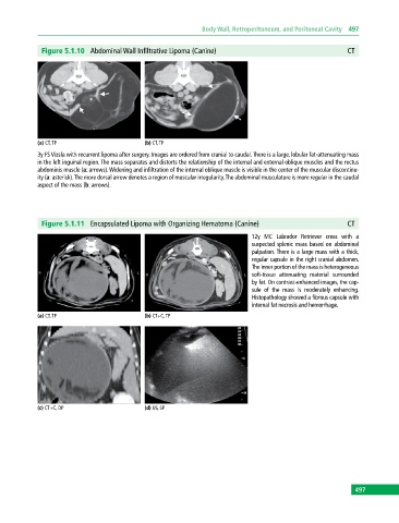
Figure 5.1.10 Abdominal Wall Infiltrative Lipoma (Canine) CT
(a) CT, TP (b) CT, TP
3y FS Vizsla with recurrent lipoma after surgery. Images are ordered from cranial to caudal. There is a large, lobular fat‐attenuating mass
in the left inguinal region. The mass separates and distorts the relationship of the internal and external oblique muscles and the rectus
abdominis muscle (a: arrows). Widening and infiltration of the internal oblique muscle is visible in the center of the muscular discontinu
ity (a: asterisk). The more dorsal arrow denotes a region of muscular irregularity. The abdominal musculature is more regular in the caudal
aspect of the mass (b: arrows).
Figure 5.1.11 Encapsulated Lipoma with Organizing Hematoma (Canine) CT
12y MC Labrador Retriever cross with a
suspected splenic mass based on abdominal
palpation. There is a large mass with a thick,
regular capsule in the right cranial abdomen.
The inner portion of the mass is heterogeneous
soft‐tissue attenuating material surrounded
by fat. On contrast‐enhanced images, the cap
sule of the mass is moderately enhancing.
Histopathology showed a fibrous capsule with
internal fat necrosis and hemorrhage.
(a) CT, TP (b) CT+C, TP
(c) CT+C, DP (d) US, SP
497