
Page 506 - Atlas of Small Animal CT and MRI
P. 506
496 Atlas of Small Animal CT and MRI
Figure 5.1.8 Retroperitoneal Abscess and Superficial Draining Tract (Canine) CT
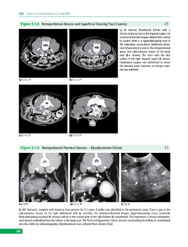
5y M German Shorthaired Pointer with a
chronic draining tract in the inguinal region. On
contrast‐enhanced images ordered from cranial
to caudal, there is a hyperattenuating tract in
the sublumbar musculature. Additional abnor
mal enhancement is seen in the retroperitoneal
space and subcutaneous tissues of the body
wall (b,c: arrows). The tract exits the skin
surface in the right inguinal region (d: arrow).
Exploratory surgery was performed to resect
the draining tracts; however, no foreign mate
rial was retrieved.
(a) CT+C, TP (b) CT+C, TP
(c) CT+C, TP (d) CT+C, TP
Figure 5.1.9 Retroperitoneal Perirenal Abscess—Mycobacterium (Feline) CT
(a) CT, TP (b) CT+C, TP (c) US, SP
8y MC Domestic Longhair with draining tract present for 2½ years. A pellet was identified in the peritoneal cavity. There is gas in the
subcutaneous tissues of the right abdominal wall (a: asterisk). On contrast‐enhanced images, hyperattenuating tissue surrounds
fluid‐attenuating material (b: arrows) lateral to the cranial pole of the right kidney (b: arrowhead). This represents a chronic retroperito
neal abscess extending from the kidney to the body wall. The fluid and hyperechoic fat (c: arrows) surrounding the kidney (c: arrowhead)
are also visible on ultrasonography. Mycobacterium was cultured from abscess fluid.
496