
Page 174 - Clinical Manual of Small Animal Endosurgery
P. 174
162 Clinical Manual of Small Animal Endosurgery
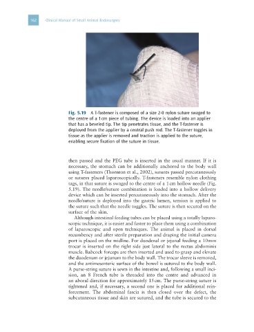
Fig. 5.19 A T-fastener is composed of a size 2-0 nylon suture swaged to
the centre of a 1 cm piece of tubing. The device is loaded into an applier
that has a beveled tip. The tip penetrates tissue, and the T-fastener is
deployed from the applier by a central push rod. The T-fastener toggles in
tissue as the applier is removed and traction is applied to the suture,
enabling secure fixation of the suture in tissue.
then passed and the PEG tube is inserted in the usual manner. If it is
necessary, the stomach can be additionally anchored to the body wall
using T-fasteners (Thornton et al., 2002), sutures passed percutaneously
or sutures placed laparoscopically. T-fasteners resemble nylon clothing
tags, in that suture is swaged to the centre of a 1 cm hollow needle (Fig.
5.19). The needle/suture combination is loaded into a hollow delivery
device which can be inserted percutaneously into the stomach. After the
needle/suture is deployed into the gastric lumen, tension is applied to
the suture such that the needle toggles. The suture is then secured on the
surface of the skin.
Although intestinal feeding tubes can be placed using a totally laparo-
scopic technique, it is easier and faster to place them using a combination
of laparoscopic and open techniques. The animal is placed in dorsal
recumbency and after sterile preparation and draping the initial camera
port is placed on the midline. For duodenal or jejunal feeding a 10 mm
trocar is inserted on the right side just lateral to the rectus abdominis
muscle. Babcock forceps are then inserted and used to grasp and elevate
the duodenum or jejunum to the body wall. The trocar sleeve is removed,
and the antimesenteric surface of the bowel is sutured to the body wall.
A purse-string suture is sewn in the intestine and, following a small inci-
sion, an 8 French tube is threaded into the centre and advanced in
an aboral direction for approximately 15 cm. The purse-string suture is
tightened and, if necessary, a second one is placed for additional rein-
forcement. The abdominal fascia is then closed over the defect, the
subcutaneous tissue and skin are sutured, and the tube is secured to the