
Page 352 - The Toxicology of Fishes
P. 352
332 The Toxicology of Fishes
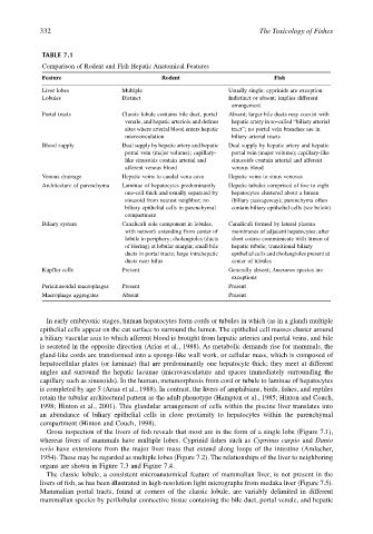
TABLE 7.1
Comparison of Rodent and Fish Hepatic Anatomical Features
Feature Rodent Fish
Liver lobes Multiple Usually single; cyprinids are exception
Lobules Distinct Indistinct or absent; implies different
arrangement
Portal tracts Classic lobule contains bile duct, portal Absent; larger bile ducts may coexist with
venule, and hepatic arteriole and defines hepatic artery in so-called “biliary arterial
sites where arterial blood enters hepatic tract”; no portal vein branches are in
microcirculation biliary arterial tracts
Blood supply Dual supply by hepatic artery and hepatic Dual supply by hepatic artery and hepatic
portal vein (major volume); capillary- portal vein (major volume); capillary-like
like sinusoids contain arterial and sinusoids contain arterial and afferent
afferent venous blood venous blood
Venous drainage Hepatic veins to caudal vena cava Hepatic veins to sinus venosus
Architecture of parenchyma Laminae of hepatocytes predominantly Hepatic tubules comprised of five to eight
one-cell thick and usually separated by hepatocytes clustered about a lumen
sinusoid from nearest neighbor; no (biliary passageway); parenchyma often
biliary epithelial cells in parenchymal contain biliary epithelial cells (see below)
compartment
Biliary system Canaliculi sole component in lobules, Canaliculi formed by lateral plasma
with network extending from center of membranes of adjacent hepatocytes; after
lobule to periphery; cholangioles (ducts short course communicate with lumen of
of Hering) at lobular margin; small bile hepatic tubule; transitional biliary
ducts in portal tracts; large intrahepatic epithelial cells and cholangioles present at
ducts near hilus center of tubules
Kupffer cells Present Generally absent; Ameiurus species are
exceptions
Perisinusoidal macrophages Present Present
Macrophage aggregates Absent Present
In early embryonic stages, human hepatocytes form cords or tubules in which (as in a gland) multiple
epithelial cells appear on the cut surface to surround the lumen. The epithelial cell masses cluster around
a biliary vascular axis to which afferent blood is brought from hepatic arteries and portal veins, and bile
is secreted in the opposite direction (Arias et al., 1988). As metabolic demands rise for mammals, the
gland-like cords are transformed into a sponge-like wall work, or cellular mass, which is composed of
hepatocellular plates (or laminae) that are predominantly one hepatocyte thick; they meet at different
angles and surround the hepatic lacunae (microvasculature and spaces immediately surrounding the
capillary such as sinusoids). In the human, metamorphosis from cord or tubule to laminae of hepatocytes
is completed by age 5 (Arias et al., 1988). In contrast, the livers of amphibians, birds, fishes, and reptiles
retain the tubular architectural pattern as the adult phenotype (Hampton et al., 1985; Hinton and Couch,
1998; Hinton et al., 2001). This glandular arrangement of cells within the piscine liver translates into
an abundance of biliary epithelial cells in close proximity to hepatocytes within the parenchymal
compartment (Hinton and Couch, 1998).
Gross inspection of the livers of fish reveals that most are in the form of a single lobe (Figure 7.1),
whereas livers of mammals have multiple lobes. Cyprinid fishes such as Cyprinus carpio and Danio
rerio have extensions from the major liver mass that extend along loops of the intestine (Amlacher,
1954). These may be regarded as multiple lobes (Figure 7.2). The relationships of the liver to neighboring
organs are shown in Figure 7.3 and Figure 7.4.
The classic lobule, a consistent microanatomical feature of mammalian liver, is not present in the
livers of fish, as has been illustrated in high-resolution light micrographs from medaka liver (Figure 7.5).
Mammalian portal tracts, found at corners of the classic lobule, are variably delimited in different
mammalian species by perilobular connective tissue containing the bile duct, portal venule, and hepatic