
Page 355 - The Toxicology of Fishes
P. 355
Liver Toxicity 335
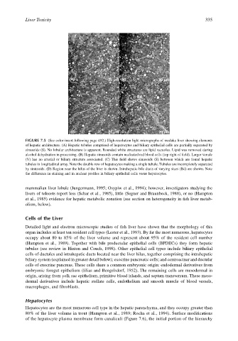
Bd
S
V
(A) 20 µm (B) 20 µm
Bd
S
S
(C) 20 µm (D) 20 µm
FIGURE 7.5 (See color insert following page 492.) High-resolution light micrographs of medaka liver showing elements
of hepatic architecture. (A) Hepatic tubules comprised of hepatocytes and biliary epithelial cells are partially separated by
sinusoids (S). No lobular architecture is apparent. Rounded white structures are lipid vacuoles. Lipid was removed during
alcohol dehydration in processing. (B) Hepatic sinusoids contain nucleated red blood cells (top right of field). Larger venule
(V) has no arterial or biliary structure associated. (C) This field shows sinusoids (S) between which are found hepatic
tubules in longitudinal array. Note the double row of hepatocytes making a single tubule. Tubules are incompletely separated
by sinusoids. (D) Region near the hilus of the liver is shown. Intrahepatic bile ducts of varying sizes (Bd) are shown. Note
the difference in staining and in nuclear profiles in biliary epithelial cells vesus hepatocytes.
mammalian liver lobule (Jungermann, 1995; Osypiw et al., 1994); however, investigators studying the
livers of teleosts report less (Schar et al., 1985), little (Segner and Braunbeck, 1988), or no (Hampton
et al., 1985) evidence for hepatic metabolic zonation (see section on heterogeneity in fish liver metab-
olism, below).
Cells of the Liver
Detailed light and electron microscopic studies of fish liver have shown that the morphology of this
organ includes at least ten resident cell types (Lester et al., 1993). By far the most numerous, hepatocytes
occupy about 80 to 85% of the liver volume and represent about 95% of the resident cell number
(Hampton et al., 1989). Together with bile preductular epithelial cells (BPDECs) they form hepatic
tubules (see review in Hinton and Couch, 1998). Other epithelial cell types include biliary epithelial
cells of ductules and intrahepatic ducts located near the liver hilus, together comprising the intrahepatic
biliary system (explained in greater detail below); exocrine pancreatic cells; and centroacinar and ductular
cells of exocrine pancreas. These cells share a common embryonic origin: endodermal derivatives from
embryonic foregut epithelium (Elias and Bengelsdorf, 1952). The remaining cells are mesodermal in
origin, arising from yolk sac epithelium, primitive blood islands, and septum transversum. These meso-
dermal derivatives include hepatic stellate cells, endothelium and smooth muscle of blood vessels,
macrophages, and fibroblasts.
Hepatocytes
Hepatocytes are the most numerous cell type in the hepatic parenchyma, and they occupy greater than
80% of the liver volume in trout (Hampton et al., 1989; Rocha et al., 1994). Surface modifications
of the hepatocyte plasma membrane form canaliculi (Figure 7.6), the initial portion of the hierarchy