
Page 356 - The Toxicology of Fishes
P. 356
336 The Toxicology of Fishes
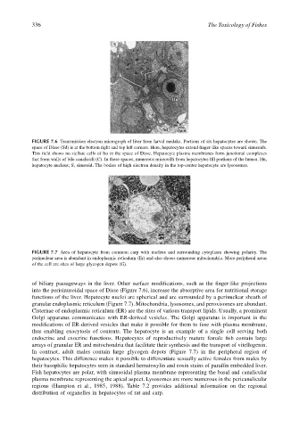
FIGURE 7.6 Transmission electron micrograph of liver from larval medaka. Portions of six hepatocytes are shown. The
space of Disse (Sd) is at the bottom right and top left corners. Here, hepatocytes extend finger-like spaces toward sinusoids.
This field shows no stellate cells of Ito in the space of Disse. Hepatocyte plasma membranes form junctional complexes
that form walls of bile canaliculi (C). In these spaces, numerous microvilli from hepatocytes fill portions of the lumen. Hn,
hepatocyte nucleus; S, sinusoid. The bodies of high electron density in the top-center hepatocyte are lysosomes.
FIGURE 7.7 Area of hepatocyte from common carp with nucleus and surrounding cytoplasm showing polarity. The
perinuclear area is abundant in endoplasmic reticulum (Er) and also shows numerous mitochondria. More peripheral areas
of the cell are sites of large glycogen depots (G).
of biliary passageways in the liver. Other surface modifications, such as the finger-like projections
into the perisinusoidal space of Disse (Figure 7.6), increase the absorptive area for nutritional storage
functions of the liver. Hepatocyte nuclei are spherical and are surrounded by a perinuclear sheath of
granular endoplasmic reticulum (Figure 7.7). Mitochondria, lysosomes, and peroxisomes are abundant.
Cisternae of endoplasmic reticulum (ER) are the sites of various transport lipids. Usually, a prominent
Golgi apparatus communicates with ER-derived vesicles. The Golgi apparatus is important in the
modifications of ER-derived vesicles that make it possible for them to fuse with plasma membrane,
thus enabling exocytosis of contents. The hepatocyte is an example of a single cell serving both
endocrine and exocrine functions. Hepatocytes of reproductively mature female fish contain large
arrays of granular ER and mitochondria that facilitate their synthesis and the transport of vitellogenin.
In contrast, adult males contain large glycogen depots (Figure 7.7) in the peripheral region of
hepatocytes. This difference makes it possible to differentiate sexually active females from males by
their basophilic hepatocytes seen in standard hematoxylin and eosin stains of paraffin embedded liver.
Fish hepatocytes are polar, with sinusoidal plasma membrane representing the basal and canalicular
plasma membrane representing the apical aspect. Lysosomes are more numerous in the pericanalicular
regions (Hampton et al., 1985, 1988). Table 7.2 provides additional information on the regional
distribution of organelles in hepatocytes of rat and carp.