
Page 354 - The Toxicology of Fishes
P. 354
334 The Toxicology of Fishes
IB
P
SV
HPV
A
L V
Ga
FIGURE 7.3 (See color insert following page 492.) Hematoxylin and eosin stain of section through adult medaka (Oryzias
latipes). Rostral structures are oriented toward the right side of the field; dorsal is top and ventral bottom. Parasaggital
section shows portions of the abdominal cavity, pericardial cavity, branchial chamber, and pharynx. A, entry to aorta; Ga,
gill arch with primary lamellae attached; HPV, hepatic portal vein in liver hilus; IB, intestinal bulb; L, liver; P, pharyngeal
mucosa with teeth; SV, sinus venosus; V, ventricle. See micron bar for magnification.
P
SV
A
HV
L
V VA
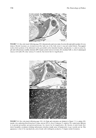
FIGURE 7.4 (See color insert following page 492.) (A) Stain and orientation are identical to Figure 7.3. A, atrium; HV,
hepatic vein conducting blood from liver to sinus venosus (SV); L, liver; P, pharynx; V, ventricle; VA, ventral aorta. Melanin
is in parietal pericardium and parietal peritoneum. See micron bar for magnification. (B). Section of adult female liver
shows basophilic cytoplasm overlying endoplasmic reticulum (purple areas of hepatocytes). Large vacuoles are fat. This
appearance is that of the reproductively active female with vitellogenin production. T, hepatic tubule formations.