
Page 360 - The Toxicology of Fishes
P. 360
340 The Toxicology of Fishes
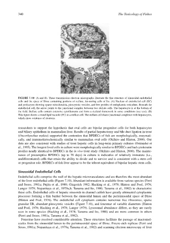
FIGURE 7.10 (A and B). These transmission electron micrographs illustrate the fine structure of sinusoidal endothelial
cells and the space of Disse containing portions of stellate, fat-storing cells of Ito. (A) Nucleus of endothelial cell (EC)
and perikaryon showing sparse mitochondria, pinocytotic vesicles, and few profiles of endoplasmic reticulum. Beneath the
endothelial cell, the arrow points to the junctional complex between two stellate cells. The hepatocyte is at the bottom of
the field. Stellate cells contain extensive cytofilaments and form a skeletal framework in some conditions (see text). (B)
This figure shows a round lipid vacuole (FC) in a stellate cell. The stellate cell shares junctional complexes with hepatocytes,
which show evidence of steatosis.
researchers to support the hypothesis that oval cells are bipolar progenitor cells for both hepatocytes
and biliary epithelium in mammalian liver. Results of partial hepatectomy and bile duct ligation in trout
(Oncorhynchus mykiss) supported the contention that BPDECs of fish are morphologically, enzymati-
cally, and immunohistochemically similar to mammalian oval cells (Okihiro and Hinton, 2000). Our
data are also consistent with studies of trout hepatic cells in long-term primary cultures (Ostrander et
al., 1995). The longest lived cells in culture were morphologically similar to BPDECs and had cytokeratin
profiles nearly identical to BPDECs in the in vivo trout study (Okihiro and Hinton, 2000). The mainte-
nance of presumptive BPDECs (up to 70 days) in culture is indicative of relatively immature (i.e.,
undifferentiated) cells that retain the ability to divide and to survive and is consistent with a stem cell
or progenitor role. BPDECs of fish liver appear to be the teleost equivalent of bipolar hepatic stem cells.
Sinusoidal Endothelial Cells
Endothelial cells comprise the wall of the hepatic microvasculature and are therefore the most abundant
of the liver endothelial cells (Figure 7.10). Abundant information is available from various species (Ferri
and Sesso, 1981a; Fujita et al., 1980; Gingerich 1982; Hacking et al., 1978; Hinton and Pool, 1976;
Langer 1979; Nopanitaya et al., 1979a,b; Tanuma and Ito, 1980; Tanuma et al., 1982) to characterize
these cells. Endothelial cells of hepatic sinusoids in channel catfish have greatly attenuated cytoplasmic
processes forming a thin barrier between the sinusoidal lumen and the perisinusoidal space of Disse
(Hinton and Pool, 1976). The endothelial cell cytoplasm contains numerous free ribosomes, sparse
granular ER, abundant pinocytotic vesicles (Figure 7.10), and fenestrae of variable diameters (Hinton
and Pool, 1976; Hacking et al., 1978; Langer 1979). Lysosomal abundance differs, as they are rarely
seen in some species (Hacking et al., 1978; Tanuma and Ito, 1980) and are more common in others
(Ferri and Sesso, 1981a; Tanuma et al., 1982).
Fenestrae have received considerable attention. These structures facilitate the passage of macromol-
ecules from the sinusoidal lumen to the perisinusoidal space of Disse. Freeze-etch replicas (Ferri and
Sesso, 1981a; Nopanitaya et al., 1979a; Tanuma et al., 1982) and scanning electron microscopy of liver