
Page 362 - The Toxicology of Fishes
P. 362
342 The Toxicology of Fishes
(A) (B)
20 µm 20 µm
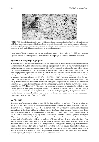
FIGURE 7.11 (See color insert following page 492.) Hematoxylin and eosin stained sections from liver of juvenile sturgeon.
(A) Melano–macrophage aggregates (black arrowheads) are seen in the connective tissue tract at margins of inflammatory
focus (eosinophilic granular leukocytes and mononuclear cells). (B) Liver parenchyma has smaller melano– macrophage
aggregates in this juvenile. Black arrowheads point to perivascular aggregates.
assessments of livers were done in these species (Hampton et al., 1989; Rocha et al., 1997), each reported
a greater number of interhepatocytic, perisinusoidal macrophages in livers of female vs. male fish.
Pigmented Macrophage Aggregates
In a recent review, the liver of mature fish was not considered to be as important to immune function
as in mammals (Rice, 2001); however, macrophage aggregates are common in the liver of certain species,
such as the sturgeon (Acipenser transmontanus) (Figure 7.11), as well as in the kidney and spleen (Agius,
1985; Kennedy-Stoskopf, 1993). The function of these cells is debated; for example, some regard these
to be the phylogenetic precursors of germinal centers, but, as others have shown, they increase in number
with age and also show an increase in number under oxidative stress. These aggregates are seen in the
presence of disease even in younger fish (Camp, 1997; Rice, 2001). In certain species of fishes, pigments
abound in these aggregates, including lipofuscin, melanin, hemosiderin, and, in some cases, ceroid (Rice,
2001). Hemosiderin is a breakdown product of hemoglobin degradation, but melanin has antioxidant
properties and is a predominant feature of macrophage aggregations in channel catfish during disease
states (Camp, 1997; Rice, 2001). Lipofuscin and ceroid are byproducts of saturated fat oxidation. Most
authors agree that macrophage aggregates are sites of inflammation, oxygen radical formation, and lipid
oxidation. In addition, the review by Rice (2001) included findings suggesting that genetic resistance to
certain diseases in channel catfish were correlated with increased numbers of splenic macrophage
aggregations (Camp, 1997; Camp et al., 2000).
Kupffer Cells
Some species of fish possess cells that resemble the fixed, resident macrophages of the mammalian liver
(Kupffer cells). Other species, despite intense investigation, seem to lack these sinusoidal lining cells
(Hampton et al., 1985; Rocha et al., 1997). Hampton et al. (1987) conducted an evaluation of resident
macrophages in the livers of brown bullhead catfish (Ictalurus nebulosus, now Ameiurus sp.). These
cells shared properties of the Kupffer cells of mammals, including phagocytosis and degradation of
erythrocytes, as well as being strongly positive for peroxidase, intensely positive for glucose-6-phosphate
dehydrogenase, and positive for phagocytosis of intravascular-induced submicron latex beads. In contrast
to mammalian Kupffer cells, resident macrophages of brown bullhead showed no invaginations of the
plasma membrane analogous to vermiform processes. Kupffer cells of mammals are very active in the
first-order defense of the liver (Cho et al., 2000; Seki et al., 2000); in the human, they perform intricate
signaling that primes other cells to react, thus providing protection against bacterial infections and
hematogenous tumor metastases (Seki et al., 2000). We do not know which cells provide this or similar
roles in fish. For those species examined, Kupffer cells are often absent, and much more information is
needed regarding this aspect of defensive roles for teleosts livers.