
Page 482 - Veterinary Toxicology, Basic and Clinical Principles, 3rd Edition
P. 482
Manganese Chapter | 30 449
VetBooks.ir 1994). Additional findings also suggest that in the absence show that Mn-induced oxidative damage and neuroinflam-
21
of extracellular Ca , Mn induces a long-lasting potentia-
mation targeted the dendritic system with profound
tion of acetylcholine (ACh) release from cardiac parasym-
dendrite regression of striatal MSNs. While a single Mn
pathetic nerve terminals following tetanic nerve exposure altered the integrity of the dendritic system and
stimulation (Kita et al., 1981). In combination with induced significant decrease in spine numbers and total
glutamate-gated cation channel activation, e.g., N-methyl- dendritic lengths of MSNs, prolonged Mn exposure led
D-aspartate receptor, secondary excitotoxicity mechanisms to further reduction in spine numbers and dendritic
play an important role in the development of Mn-induced lengths (Milatovicetal.,2009). In essence, MSNs neurode-
neurodegeneration. generation could result from loss of spines, removing
Neurotoxicity of Mn reflects alterations in the integrity the pharmacological target for DA-replacement therapy,
of DAergic striatal neurons and DA neurochemistry, without overt MSNs death (Stephens et al., 2005; Zaja-
including decreased DA transport function and/or striatal Milatovic et al., 2005).
DA levels. The striatum is a major recipient structure of
neuronal afferents in the basal ganglia. It receives excit- TOXICITY
atory input from the cortex and DAergic input from sub-
stantia nigra and projects to the internal segment of the Mn is considered to be one of the least toxic of the
globus pallidus (Dimova et al., 1993; Saka et al., 2002). essential elements (NRC, 2005). There are no reports of
Nigrostriatal DAergic neurons appear to be particularly acute toxicity of Mn in animals. Therefore, all toxicity
sensitive to Mn-induced toxicity (Sloot and Gramsbergen, studies described here are chronic in nature. A diet can be
1994; Sloot et al., 1994; Defazio et al., 1996). Intense or consumed without any adverse effect when the Mn level
prolonged Mn exposure in adulthood causes long-term is 2000 ppm for calves, 3000 ppm for sheep, 3000 ppm
reductions in striatal DA levels and induces a loss of auto- for chickens, 4000 ppm for turkeys and 7000 ppm
receptor control over DA release (Autissier et al., 1982; for rats. However, decreased growth is observed at
Komura and Sakamoto, 1992). Nigrostriatal DA axons 500 3000 ppm in swine. These data indicate that pigs are
synapse onto striatal medium spiny neurons (MSNs), and more sensitive to excess Mn than other livestock (NRC,
these neurons have radially projecting dendrites that are 2005). Mn at a 5000 ppm dietary level is lethal to preru-
densely studded with spines (Wilson and Groves, 1980). minant calves (Puls, 1994). Clinical signs of toxicity
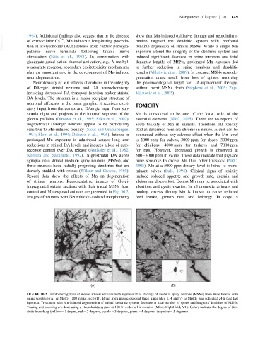
Recent data show the effects of Mn on degeneration include reduced appetite and growth rate, anemia and
of striatal neurons. Representative images of Golgi- abdominal discomfort. Excess Mn may be associated with
impregnated striatal sections with their traced MSNs from abortions and cystic ovaries. In all domestic animals and
control and Mn-exposed animals are presented in Fig. 30.2. poultry, excess dietary Mn is known to cause reduced
Images of neurons with Neurolucida-assisted morphometry feed intake, growth rate, and lethargy. In dogs, a
(A) (B)
FIGURE 30.2 Photomicrographs of mouse striatal sections with representative tracings of medium spiny neurons (MSNs) from mice treated with
saline (control) (A) or MnCl 2 (100 mg/kg, s.c.) (B). Brain from mouse exposed three times (day 1, 4 and 7) to MnCl 2 was collected 24 h post last
injection. Treatment with Mn-induced degeneration of striatal dendritic system, decrease in total number of spines and length of dendrites of MSNs.
Tracing and counting are done using a Neurolucida system at 100 3 under oil immersion (MicroBrightField, VT). Colors indicate the degree of den-
dritic branching (yellow 5 1 degree, red 5 2 degrees, purple 5 3 degrees, green 5 4 degrees, turquoise 5 5 degrees).