Page 291 - Withrow and MacEwen's Small Animal Clinical Oncology, 6th Edition
P. 291
270   PART III    Therapeutic Modalities for the Cancer Patient


         enhance apoptosis in tumor and endothelial cells, 370,374–376    before a standard dose of DOX in tumor-bearing dogs, at dosages
         and directly inhibit tumor cell proliferation. 375,377–379  Consis-  sufficient to enhance histone acetylation in tumor and periph-
                                                               eral blood mononuclear cells, but not associated with toxicity or
         tent with its role as a transcriptional repressor, inhibition of
  VetBooks.ir  HDACs has been shown to induce differentiation in thyroid     apparent potentiation of DOX’s adverse effects. 392
                                                       380–384
         and prostate cancer, neuroblastoma, and the leukemias.
         HDAC inhibition enhances the in  vitro and in  vivo efficacy   The Proteasome
         of multiple standard cytotoxic therapies, antibodies and small
         molecules. 367,370,376,385–393                        The abundance of cellular proteins is tightly controlled at the
            Two HDAC inhibitors, vorinostat (SAHA, Zolinza,    levels of both production and destruction. Protein production
         Merck) and romidepsin (FK228, Istodax, Celgene), have been   can be modified at the transcriptional and posttranscriptional
         approved by the FDA for the treatment of cutaneous T-cell lym-  level, and therapies based on these approaches are relatively abun-
         phoma, 394–396  and  a large number  of additional  agents are in   dant. Until relatively recently, little attention had been paid to
         intermediate- to late-stage clinical trials. A number of studies   the manipulation of protein degradation as a therapeutic modal-
         are being conducted evaluating HDAC inhibitors for various   ity. The ubiquitin–proteasome pathway (UPP) is responsible for
         other hematologic and solid tumors, and early studies combin-  the degradation of the majority of intracellular proteins, and is
         ing these drugs with other targeted and cytotoxic therapies have   responsible for the regulation of many proteins with key roles in
         been reported. 397–400                                cancer.
            Recent in vitro studies have demonstrated potent induction   The 26S proteasome is a large multiprotein complex contain-
         of histone acetylation, growth inhibition, and induction of apop-  ing ubiquitin recognition domains, which bind ubiquitinated
         tosis in a variety of canine tumor cell lines treated with either   proteins tagged for degradation, and proteolytic domains with
         vorinostat or the novel HDAC inhibtior OSU-HDAC42 (AR-  trypsin-like, chymotrypsin-like, and caspase-like activity, which
         42)  401,402 ; and inhibition of canine prostate cancer metastasis to   degrade proteins into short polypeptide sequences. 405  It is respon-
         bone was observed after AR-42 treatment in a murine xenograft   sible for the degradation of a variety of proteins responsible for
         model. 403  Synergy between vorinostat and the DNMT inhibi-  cell-cycle regulation, angiogenesis, apoptosis, and chemotherapy
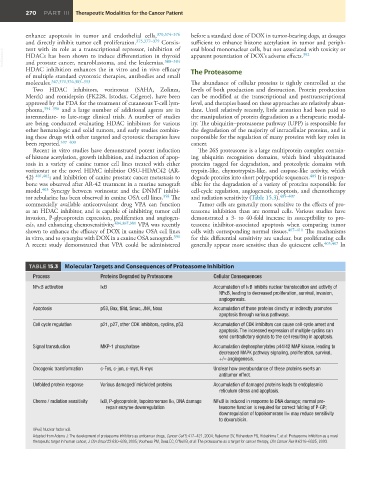
         tor zebularine has been observed in canine OSA cell lines. 358  The   and radiation sensitivity (Table 15.3). 405–407
         commercially available anticonvulsant drug  VPA can function   Tumor cells are generally more sensitive to the effects of pro-
         as an HDAC inhibitor, and is capable of inhibiting tumor cell   teasome inhibition than are normal cells.  Various studies have
         invasion, P-glycoprotein expression, proliferation and angiogen-  demonstrated a 3- to 40-fold increase in susceptibility to pro-
         esis, and enhancing chemosensitivity. 404,367,381  VPA was recently   teasome  inhibitor-associated  apoptosis  when  comparing  tumor
         shown to enhance the efficacy of DOX in canine OSA cell lines   cells with corresponding normal tissues. 407–411  The mechanisms
         in vitro, and to synergize with DOX in a canine OSA xenograft. 391    for this differential sensitivity are unclear, but proliferating cells
         A recent study demonstrated that  VPA could be administered   generally appear more sensitive than do quiescent cells. 405,407  In


          TABLE 15.3     Molecular Targets and Consequences of Proteasome Inhibition
            Process               Proteins Degraded by Proteasome      Cellular Consequences
            NFκB activation       IκB                                  Accumulation of IκB inhibits nuclear translocation and activity of
                                                                         NFκB, leading to decreased proliferation, survival, invasion,
                                                                         angiogenesis.
            Apoptosis             p53, Bax, tBid, Smac, JNK, Noxa      Accumulation of these proteins directly or indirectly promotes
                                                                         apoptosis through various pathways.
            Cell cycle regulation  p21, p27, other CDK inhibitors, cyclins, p53  Accumulation of CDK inhibitors can cause cell-cycle arrest and
                                                                         apoptosis. The increased expression of multiple cyclins can
                                                                         send contradictory signals to the cell resulting in apoptosis.
            Signal transduction   MKP-1 phosphatase                    Accumulation dephosphorylates p44/42 MAP kinase, leading to
                                                                         decreased MAPK pathway signaling, proliferation, survival,
                                                                         +/– angiogenesis.
            Oncogenic transformation  c-Fos, c-jun, c-myc, N-myc       Unclear how overabundance of these proteins exerts an
                                                                           antitumor effect.
            Unfolded protein response  Various damaged/ misfolded proteins  Accumulation of damaged proteins leads to endoplasmic
                                                                           reticulum stress and apoptosis.
            Chemo / radiation sensitivity  IκB, P-glycoprotein, topoisomerase IIα, DNA damage   NFκB is induced in response to DNA damage; normal pro-
                                    repair enzyme downregulation         teasome function is required for correct folding of P-GP;
                                                                         downregulation of topoisomerase IIα may reduce sensitivity
                                                                         to doxorubicin.
            NFκB, Nuclear factor κB.
            Adapted from Adams J: The development of proteasome inhibitors as anticancer drugs, Cancer Cell 5:417–421, 2004; Rajkumar SV, Richardson PG, Hideshima T, et al: Proteasome inhibition as a novel
            therapeutic target in human cancer, J Clin Oncol 23:630–639, 2005; Voorhees PM, Dees EC, O’Neil B, et al: The proteasome as a target for cancer therapy, Clin Cancer Res 9:6316–6325, 2003.

