
Page 456 - Withrow and MacEwen's Small Animal Clinical Oncology, 6th Edition
P. 456
434 PART IV Specific Malignancies in the Small Animal Patient
TABLE 23.2 Clinical Staging (TNM) of Oral Tumors in
Dogs and Cats 70
VetBooks.ir Clinical Staging System for Oral Tumors
Primary Tumor (T)
Tis Tumor in situ
T1 Tumor <2 cm in diameter at greatest dimension
T1a Without evidence of bone invasion
T1b With evidence of bone invasion
T2 Tumor 2–4 cm in diameter at greatest dimension
T2a Without evidence of bone invasion
T2b With evidence of bone invasion
T3 Tumor >4 cm in diameter at greatest dimension
T3a Without evidence of bone invasion
T3b With evidence of bone invasion
Regional Lymph Nodes (N)
A N0 No regional lymph node metastasis
N1 Movable ipsilateral lymph nodes
N1a No evidence of lymph node metastasis
N1b Evidence of lymph node metastasis
N2 Movable contralateral lymph nodes
N2a No evidence of lymph node metastasis
N2b Evidence of lymph node metastasis
N3 Fixed lymph nodes
Distant Metastasis (M)
M0 No distant metastasis
M1 Distant metastasis [specify site(s)]
Stage
Grouping Tumor (T) Nodes (N) Metastasis (M)
I T1 N0, N1a, N2a M0
II T2 N0, N1a, N2a M0
B III T3 N0, N1a, N2a M0
IV Any T N1b M0
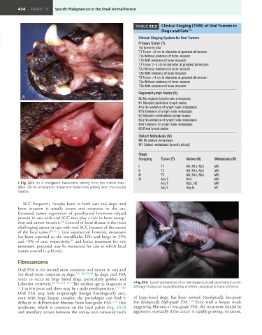
• Fig. 23.1 (A) A malignant melanoma arising from the rostral man- Any T N2b, N3 M0
dible. (B) An amelanotic malignant melanoma arising from the caudal Any T Any N M1
maxilla.
SCC frequently invades bone in both cats and dogs, and
bone invasion is usually severe and extensive in the cat.
Increased tumor expression of parathyroid hormone–related
protein in cats with oral SCC may play a role in bone resorp-
tion and tumor invasion. Control of local disease is the most
88
challenging aspect in cats with oral SCC because of the extent
of the local tumor 89–112 ; [not superscript] however, metastasis
has been reported to the mandibular LNs and lungs in 31%
and 10% of cats, respectively, and hence treatment for this
95
metastatic potential may be warranted for cats in which local
tumor control is achieved.
Fibrosarcoma
Oral FSA is the second most common oral tumor in cats and
the third most common in dogs. 2,5, 20–24,90 In dogs, oral FSA
tends to occur in large breed dogs, particularly golden and
Labrador retrievers. 20–24,113–117 The median age at diagnosis is • Fig. 23.2 Typical appearance of an oral squamous cell carcinoma in a cat.
7.3 to 8.6 years and there may be a male predisposition. 113–116 Although these can be proliferative and firm, ulceration is more common.
Oral FSA may look surprisingly benign histologically and,
even with large biopsy samples, the pathologist can find it of large-breed dogs, has been termed histologically low-grade
difficult to differentiate fibroma from low-grade FSA. 113 This but biologically high-grade FSA. 113 Even with a biopsy result
syndrome, which is common on the hard palate (Fig. 23.3) suggesting fibroma or low-grade FSA, the treatment should be
and maxillary arcade between the canine and carnassial teeth aggressive, especially if the cancer is rapidly growing, recurrent,