
Page 460 - Withrow and MacEwen's Small Animal Clinical Oncology, 6th Edition
P. 460
VetBooks.ir
A B
C D
E
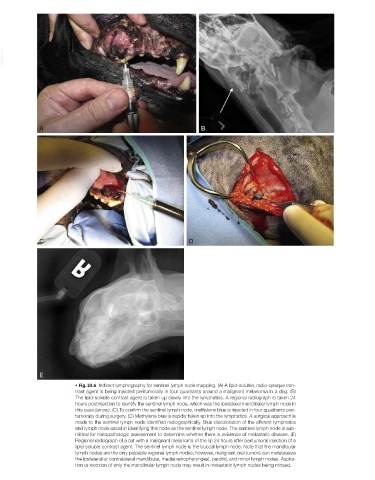
• Fig. 23.6 Indirect lymphography for sentinel lymph node mapping. (A) A lipid-soluble, radio-opaque con-
trast agent is being injected peritumorally in four quadrants around a malignant melanoma in a dog. (B)
The lipid-soluble contrast agent is taken up slowly into the lymphatics. A regional radiograph is taken 24
hours postinjection to identify the sentinel lymph node, which was the ipsilateral mandibular lymph node in
this case (arrow). (C) To confirm the sentinel lymph node, methylene blue is injected in four quadrants peri-
tumorally during surgery. (D) Methylene blue is rapidly taken up into the lymphatics. A surgical approach is
made to the sentinel lymph node identified radiographically. Blue discoloration of the afferent lymphatics
and lymph node assist in identifying this node as the sentinel lymph node. The sentinel lymph node is sub-
mitted for histopathologic assessment to determine whether there is evidence of metastatic disease. (E)
Regional radiograph of a cat with a malignant melanoma of the lip 24 hours after peritumoral injection of a
lipid-soluble contrast agent. The sentinel lymph node is the buccal lymph node. Note that the mandibular
lymph nodes are the only palpable regional lymph nodes; however, malignant oral tumors can metastasize
the ipsilateral or contralateral mandibular, medial retropharyngeal, parotid, and minor lymph nodes. Aspira-
tion or excision of only the mandibular lymph node may result in metastatic lymph nodes being missed.