
Page 457 - Withrow and MacEwen's Small Animal Clinical Oncology, 6th Edition
P. 457
CHAPTER 23 Cancer of the Gastrointestinal Tract 435
VetBooks.ir
• Fig. 23.3 Typical appearance of a biologically high-grade but histologi-
cally low-grade fibrosarcoma. These often appear histologically benign or
low-grade, but have an aggressive local behavior. Wide surgical resection
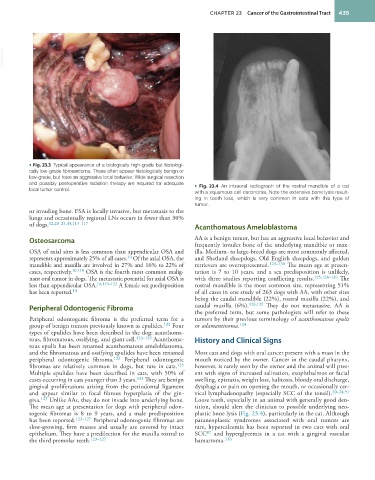
and possibly postoperative radiation therapy are required for adequate • Fig. 23.4 An intraoral radiograph of the rostral mandible of a cat
local tumor control. with a squamous cell carcinoma. Note the extensive bone lysis result-
ing in tooth loss, which is very common in cats with this type of
tumor.
or invading bone. FSA is locally invasive, but metastasis to the
lungs and occasionally regional LNs occurs in fewer than 30%
of dogs. 12,20–24,43,113–117 Acanthomatous Ameloblastoma
Osteosarcoma AA is a benign tumor, but has an aggressive local behavior and
frequently invades bone of the underlying mandible or max-
OSA of axial sites is less common than appendicular OSA and illa. Medium- to large-breed dogs are most commonly affected,
represents approximately 25% of all cases. Of the axial OSA, the and Shetland sheepdogs, Old English sheepdogs, and golden
10
mandible and maxilla are involved in 27% and 16% to 22% of retrievers are overrepresented. 123–130 The mean age at presen-
cases, respectively. 10,118 OSA is the fourth most common malig- tation is 7 to 10 years, and a sex predisposition is unlikely,
nant oral tumor in dogs. The metastatic potential for axial OSA is with three studies reporting conflicting results. 125,128–130 The
less than appendicular OSA. 10,119–122 A female sex predisposition rostral mandible is the most common site, representing 51%
10
has been reported. of all cases in one study of 263 dogs with AA, with other sites
being the caudal mandible (22%), rostral maxilla (22%), and
Peripheral Odontogenic Fibroma caudal maxilla (6%). 128,129 They do not metastasize. AA is
the preferred term, but some pathologists will refer to these
Peripheral odontogenic fibroma is the preferred term for a tumors by their previous terminology of acanthomatous epulis
group of benign tumors previously known as epulides. 123 Four or adamantinoma. 124
types of epulides have been described in the dog: acanthoma-
tous, fibromatous, ossifying, and giant cell. 123–135 Acanthoma- History and Clinical Signs
tous epulis has been renamed acanthomatous ameloblastoma,
and the fibromatous and ossifying epulides have been renamed Most cats and dogs with oral cancer present with a mass in the
peripheral odontogenic fibroma. 123 Peripheral odontogenic mouth noticed by the owner. Cancer in the caudal pharynx,
fibromas are relatively common in dogs, but rare in cats. 135 however, is rarely seen by the owner and the animal will pres-
Multiple epulides have been described in cats, with 50% of ent with signs of increased salivation, exophthalmos or facial
cases occurring in cats younger than 3 years. 135 They are benign swelling, epistaxis, weight loss, halitosis, bloody oral discharge,
gingival proliferations arising from the periodontal ligament dysphagia or pain on opening the mouth, or occasionally cer-
and appear similar to focal fibrous hyperplasia of the gin- vical lymphadenopathy (especially SCC of the tonsil). 20–24,91
giva. 123 Unlike AAs, they do not invade into underlying bone. Loose teeth, especially in an animal with generally good den-
The mean age at presentation for dogs with peripheral odon- tition, should alert the clinician to possible underlying neo-
togenic fibromas is 8 to 9 years, and a male predisposition plastic bone lysis (Fig. 23.4), particularly in the cat. Although
has been reported. 123–127 Peripheral odontogenic fibromas are paraneoplastic syndromes associated with oral tumors are
slow-growing, firm masses and usually are covered by intact rare, hypercalcemia has been reported in two cats with oral
87
epithelium. They have a predilection for the maxilla rostral to SCC and hyperglycemia in a cat with a gingival vascular
the third premolar teeth. 123–127 hamartoma. 136 