
Page 458 - Withrow and MacEwen's Small Animal Clinical Oncology, 6th Edition
P. 458
436 PART IV Specific Malignancies in the Small Animal Patient
Diagnostic Techniques and Workup
VetBooks.ir The diagnosis and clinical staging of animals with oropharyngeal
masses is imperative before definitive surgical excision. A biopsy
is required for definitive diagnosis and this will assist the clinician
in determining biologic behavior and prognosis. Clinical staging
consists of evaluating the extent of the local tumor and the pres-
ence of metastatic disease. The regional LNs and lungs are the two
most common sites of metastasis in cats and dogs with oral tum
ors. 30–68,76–85,89–122 The procedures required for the diagnosis and
clinical staging of animals with oral cancer can usually be per-
formed under a short general anesthesia.
Diagnosis
A large incisional biopsy is often required for a definitive diag-
nosis. Fine-needle aspirate (FNA) or impression smear cytology
has traditionally been considered unrewarding because many oral
tumors are associated with a high degree of necrosis and inflam-
mation; however, one prospective study of 114 cats and dogs with
oral masses showed that, in comparison to definitive histopatho-
logic results, FNA cytology had a diagnostic accuracy rate of 98%
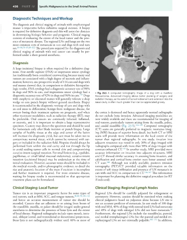
in dogs and 96% in cats, and impressions smear cytology had a • Fig. 23.5 A computed tomography image of a dog with a maxillary
diagnostic accuracy rate of 92% in dogs and 96% in cats. 137 Dogs fibrosarcoma. Advanced imaging allows better planning of surgery and
with exophytic or ulcerated masses will generally tolerate a deep radiation therapy, as the extent of bone involvement and extension into the
wedge or core punch biopsy without general anesthesia. Biopsy nasal cavity is often much greater than can be appreciated grossly.
is recommended in the diagnostic workup of cats and dogs with
an oral mass to differentiate benign from malignant disease, for
owners basing their treatment options on prognosis, and when the cortex is destroyed and hence apparently normal radiographs
other treatment modalities, such as radiation therapy (RT), may do not exclude bone invasion. Advanced imaging modalities are
be preferable. Oral cancers are commonly infected, inflamed, now widely available and these are recommended for imaging of
or necrotic, and it is important to obtain a large representative oral tumors, particularly tumors arising from the maxilla, palate,
specimen. Cautery may distort the specimen and should be used and caudal mandible (Fig. 23.5). 143–145 Computed tomography
for hemostasis only after blade incision or punch biopsy. Large (CT) scans are generally preferred to magnetic resonance imag-
samples of healthy tissue at the edge and center of the lesion ing (MRI) because of superior bone detail, but both CT or MRI
will increase the diagnostic yield, but care must be taken not to scans will provide more information on the local extent of the
contaminate normal tissue, which cannot be removed with sur- tumor than regional radiographs. In one study, invasion into
gery or included in the radiation field. Biopsies should always be adjacent structures was noted in only 30% of dogs imaged with
performed from within the oral cavity and not through the lip radiographs compared with more than 90% of dogs imaged with
to avoid seeding tumor cells in normal skin and compromising contrast-enhanced CT. 145 In another study, MRI provided more
curative-intent surgical resection. For small lesions (e.g., epulides, accurate information on invasion into adjacent structures, MRI
papillomas, or small labial mucosal melanoma), curative-intent and CT showed similar accuracy in assessing bone invasion, and
resection (excisional biopsy) may be undertaken at the time of calcification and cortical bone erosion were better assessed with
initial evaluation. However, accurate notes should be included in CT scan. 144 Although not widely available, positron emission
the medical records, and/or photographic evidence, to detail the tomography (PET)/CT provided valuable information on the
size and anatomic location of the mass if excision is incomplete extent of soft tissue infiltration and presence of LN metastasis in
and further treatment is required. For more extensive disease, cats with oral SCC in comparison to CT. 146,147 This information
waiting for biopsy results is recommended so that appropriate is important for planning the definitive surgical procedure (or RT
treatment plans can be formulated. if indicated).
Clinical Staging: Local Tumor Clinical Staging: Regional Lymph Nodes
Tumor size is an important prognostic factor for some types of Regional LNs should be carefully palpated for enlargement or
oral tumors, such as MM, SCC, and tongue tumors, 40,41,78,138–142 asymmetry. However, caution should be exercised when making
and hence an accurate measurement of tumor size should be clinical judgments based on palpation alone because LN size is
recorded. Cancers that are adherent to or arising from bones of not an accurate predictor of metastasis. In one study of 100 dogs
the mandible, maxilla, or palate should be imaged under general with oral MM, 40% of dogs with normal sized LNs had metastasis
34
anesthesia to determine the presence of bone lysis and the extent and 49% of dogs with enlarged LNs did not have metastasis.
of local disease. Regional radiographs include open mouth, intra- Furthermore, the regional LNs include the mandibular, parotid,
oral, oblique lateral, and ventrodorsal or dorsoventral projections. and medial retropharyngeal LNs; but the parotid and medial ret-
Bone lysis is not radiographically evident until 40% or more of ropharyngeal LNs are not externally palpable. 148–151 In addition,