
Page 305 - Veterinary Histology of Domestic Mammals and Birds, 5th Edition
P. 305
Male reproductive system (organa genitalia masculina) 287
VetBooks.ir
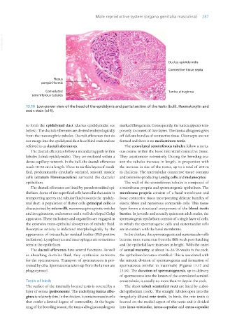
13.16 Low-power view of the head of the epididymis and partial section of the testis (bull). Haematoxylin and
eosin stain (x14).
to form the epididymal duct (ductus epididymidis; see marked fibrogenesis. Consequently, the tunica appears tem-
below). The ductuli efferentes are derived embryologically porarily to consist of two layers. The tunica albuginea gives
from the mesonephric tubules. Ductuli efferentes that do off delicate bundles of connective tissue. Clear septa are not
not merge into the epididymal duct have blind ends and are formed and there is no mediastinum testis.
referred to as ductuli aberrantes. The convoluted seminiferous tubules follow a tortu-
The ductuli efferentes follow a meandering path within ous course within the loose interstitial connective tissue.
lobules (lobuli epididymidis). They are enclosed within a They anastomose extensively. During the breeding sea-
dense capillary network. In the bull, the ductuli efferentes son the tubules increase in length, in proportion with
reach 50–80 cm in length. Three to six thin layers of modi- the increase in size of the testes, up to a total of 250 m
fied, predominantly circularly oriented, smooth muscle in chickens. The intertubular connective tissue contains
cells (stratum fibromusculare) surround the ductular androstenone-producing Leydig cells and melanocytes.
epithelium. The wall of the seminiferous tubules is composed of
The ductuli efferentes are lined by pseudostratified epi- a membrana propria and spermatogenic epithelium. The
thelium. Some of the superficial cells have cilia that assist in membrana propria consists of a basal membrane and
transporting sperm and tubular fluid towards the epididy- loose connective tissue incorporating delicate bundles of
mal duct. A population of flatter cells (principal cells) is elastic fibres and numerous contractile cells. This tissue
characterised by microvilli, numerous pinocytotic vesicles layer forms a structural component of the blood–testis
and invaginations, endosomes and a well-developed Golgi barrier. In juvenile and sexually quiescent adult males, the
apparatus. These inclusions and organelles are engaged in spermatogenic epithelium consists of a single layer of cells,
the extensive trans-epithelial absorption of tubular fluid. in which the spermatogenic cells and sustentacular cells
Resorptive activity is indicated morphologically by the are in contact with the basal membrane.
appearance of intracellular residual bodies (PAS-positive In the chicken, the spermatogenic and sustentacular cells
inclusions). Lymphocytes and macrophages are sometimes become more numerous from the fifth week post-hatching
seen in the epithelium. and the epithelial layer increases in height. With the onset
The ductuli efferentes have several functions. As well of sexual maturity, at about 16–24/26 weeks in the cock,
as absorbing ductular fluid, they synthesise nutrients the epithelium becomes stratified. This is associated with
for the spermatozoa. Transport of spermatozoa is pro- the mitotic division of spermatogonia and formation of
moted by cilia. Spermatozoa taken up from the lumen are spermatozoa (similar to mammals) (Figures 13.17 and
phagocytosed. 13.18). The duration of spermatogenesis, up to delivery
of spermatozoa into the lumen of the convoluted seminif-
Testis of birds erous tubule, is usually no more than 12 days in the cock.
The surface of the internally located testis is covered by a The short tubuli seminiferi recti are lined by cuboi-
layer of serosa (peritoneum). The underlying tunica albu- dal epithelium (cock). The straight tubules open into the
ginea is relatively thin. In the chicken, it contains muscle cells irregularly dilated rete testis. In birds, the rete testis is
that confer a limited degree of contractility. At the begin- located on the medial aspect of the testis and is divided
ning of the breeding season, the tunica albuginea undergoes into intra-testicular, intra-capsular and extra-capsular
Vet Histology.indb 287 16/07/2019 15:04