Page 304 - Veterinary Histology of Domestic Mammals and Birds, 5th Edition
P. 304
286  Veterinary Histology of Domestic Mammals and Birds


                   germ cells. Cytoplasmic streaming results in the direction  Straight testicular tubules
       VetBooks.ir  spermatozoa into the tubular lumen (spermiation) is a  The convoluted seminiferous tubules are continued by
                   of spermatocytes towards the tubular lumen. Release of  (tubuli seminiferi recti)

                   complex process that commences with dissolution of the  short, straight testicular tubules (tubuli seminiferi recti)
                   tubulobulbar complexes between the sustentacular cell  which open into the rete testis. The straight testicular
                   and the head of the spermatozoon (Figure 13.13).  tubules are usually short, though some are elongated in
                      Secretion of tubular fluid components: Tubular fluid  the boar and stallion. In the dog and bull, groups of susten-
                   is produced in large quantities. Like intracellular fluid, it is  tacular cells form a club-shaped intraluminal projection at
                   rich in potassium. The tubular fluid carries spermatozoa  the transition from the convoluted to the straight tubules
                   through the ductuli efferentes into the ductus epididymidis  (terminal segment). These contribute to phagocytosis and
                   (epididymal duct), where they complete their maturation.  prevent retrograde flow of tubular fluid.
                      Formation of the blood–testis barrier: The blood–tes-  The straight testicular tubules are lined with simple
                   tis barrier separates the circulatory system (blood and  cuboidal to columnar resorptive epithelium containing
                   lymph) and the interior of the convoluted seminiferous  lymphocytes and macrophages.
                   tubules. While in common usage, the term ‘blood–testis
                   barrier’ is misleading. It consists of the tight seal formed   Rete testis
                   between the plasmalemma of neighbouring sustentacular  The rete testis consists of anastomosing straight tubules.
                   cells by basolateral intercellular junctions. This gives rise  These are surrounded by the loose connective tissue and
                   to two compartments within the tubular epithelium:  elastic meshwork of the mediastinum testis, and by myo-
                                                                  fibroblasts (Figures 13.3 and 13.14). The epithelium of the
                     ·  a basal compartment containing spermatogonia and  rete testis is simple squamous to columnar. Two epithe-
                     ·  an adluminal compartment containing spermato-  lial layers are present in the bull. The epithelium is highly
                       cytes and spermatids (Figure 13.11).       secretory and increases the volume of the tubular fluid.
                                                                  Androgens in the tubular fluid are absorbed across the
                   In the adluminal compartment, the sustentacular cells  epithelium to a limited extent.
                   provide protection for the spermatocytes and spermatids.
                   Maintenance of the barrier is important for the secre-  Ductuli efferentes testis
                   tory functions of the sustentacular cells, through which  At the extremitas capitata of the testis, the ductuli efferen-
                   an osmotic gradient is established in the luminal region,  ties testis penetrate the tunica albuginea and pass into the
                   and for maintenance of an optimal environment for sper-  head of the epididymis (Figures 13–2 and 13–3 to 13–16).
                   miogenesis. The vulnerable spermatids are protected from  A species-dependent number of ductuli efferentes (stal-
                   autoimmune reactions, toxins and mutagens.     lion 12–23, bull 12–13, boar 14–21, dog 15–16) combines


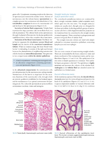
                   13.14  Testis (bull). Haematoxylin and eosin stain (x30).  13.15  Ductuli efferentes of the epididymis (stal-
                                                                  lion). The lumen contains agglutinated spermatozoa.
                                                                  Haematoxylin and eosin stain (x100).


       Vet Histology.indb   286                                                                                  16/07/2019   15:04

