Page 306 - Veterinary Histology of Domestic Mammals and Birds, 5th Edition
P. 306
288  Veterinary Histology of Domestic Mammals and Birds


       VetBooks.ir


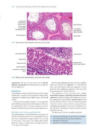
                   13.17  Testis (cock). Haematoxylin and eosin stain (x120).


                   13.18  Testis (cock). Haematoxylin and eosin stain (x440).


                   components. The last of these opens into the ductuli   Based on its morphological features, the ductus epididy-
                   efferentes proximales from which sperm are conducted  midis can be divided into segments (1–6 in the bull and
                   into the epididymis.                           ram), each with distinctive functions. Segments 1–3 lie in
                                                                  the head of the epididymis, segments 4 and 5 are in the
                   Epididymis                                     body and segment 6 is found in the tail.
                   The epididymis is firmly attached to the testis at the margo   The epithelium of the epididymal duct is  pseu-
                   epididymalis. It has three macroscopically distinguishable  dostratified columnar (epithelium pseudostratificatum
                   components: the head (caput), body (corpus) and the tail  columnare). Distally the height of the epithelium decreases.
                   (cauda).                                       The epithelium is surrounded by smooth muscle which
                      The head of the epididymis (Figure 13.16) contains the  becomes distinctly thicker in the tail. Adjacent segments
                   ductuli efferentes testis and the initial segment of the duc-  of the duct wall are connected by loose connective tissue
                   tus epididymidis (refer to Veterinary Anatomy of  Domestic  containing macrophages, leucocytes and abundant ves-
                   Mammals: Textbook and Colour Atlas for more information).  sels and nerves. The density of capillary bundles increases
                                                                  markedly in the tail.
                   Epididymal duct (ductus epididymidis)             The functions of the ductus epididymis include:
                   The epididymal duct is formed from the convergence of
                   the ductuli efferentes testis. It is tremendously tortuous   ·  maturation and storage of spermatozoa, and deliv-
                   and varies considerably in length (stallion 72–81 m, bull   ery of spermatozoa into the ductus deferens,
                   40–50 m, boar 17–18 m, dog 5–8 m). Despite this, the time   ·  absorption of fluid and
                   taken for transport of spermatozoa through the duct is   ·  secretion of metabolically active substances.
                   10–15 days in all mammals.


       Vet Histology.indb   288                                                                                  16/07/2019   15:04

