
Page 146 - Feline diagnostic imaging
P. 146
8.5 rbit 145
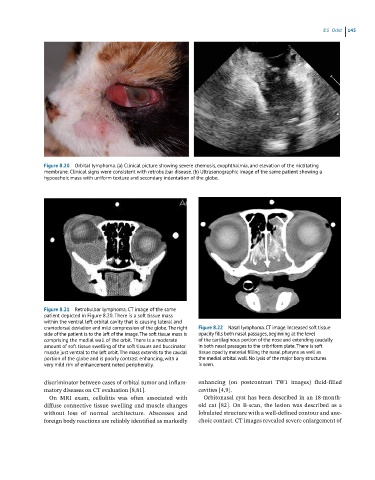
Figure 8.20 Orbital lymphoma. (a) Clinical picture showing severe chemosis, exophthalmia, and elevation of the nictitating
membrane. Clinical signs were consistent with retrobulbar disease. (b) Ultrasonographic image of the same patient showing a
hypoechoic mass with uniform texture and secondary indentation of the globe.
Figure 8.21 Retrobulbar lymphoma. CT image of the same
patient depicted in Figure 8.20. There is a soft tissue mass
within the ventral left orbital cavity that is causing lateral and
craniodorsal deviation and mild compression of the globe. The right Figure 8.22 Nasal lymphoma. CT image. Increased soft tissue
side of the patient is to the left of the image. The soft tissue mass is opacity fills both nasal passages, beginning at the level
comprising the medial wall of the orbit. There is a moderate of the cartilaginous portion of the nose and extending caudally
amount of soft tissue swelling of the soft tissues and buccinator in both nasal passages to the cribriform plate. There is soft
muscle just ventral to the left orbit. The mass extends to the caudal tissue opacity material filling the nasal pharynx as well as
portion of the globe and is poorly contrast enhancing, with a the medial orbital wall. No lysis of the major bony structures
very mild rim of enhancement noted peripherally. is seen.
discriminator between cases of orbital tumor and inflam- enhancing (on postcontrast TW1 images) fluid‐filled
matory diseases on CT evaluation [8,81]. cavities [4,9].
On MRI exam, cellulitis was often associated with Orbitonasal cyst has been described in an 18‐month‐
diffuse connective tissue swelling and muscle changes old cat [82]. On B‐scan, the lesion was described as a
without loss of normal architecture. Abscesses and lobulated structure with a well‐defined contour and ane -
foreign body reactions are reliably identified as markedly choic contact. CT images revealed severe enlargement of