
Page 142 - Feline diagnostic imaging
P. 142
8.5 rbit 141
It can be difficult to distinguish a RD from organized
vitreal strands and membranes [4,7,38]. Vitreal mem-
brane is usually not attached to the optic disc, and there is
weaker echogenicity, less even thickness, and greater
mobility than RD. By decreasing the gain, the vitreal
membranous echo will disappear sooner than the echo of
RD [10,12,19,38,65,66].
In one study, misdiagnosis between vitreal mem-
branes and RD using B‐mode ultrasound was observed
in 18.2% of cases in dogs and cats. In 91% of these cases,
the use of color Doppler imaging was unsuccessful in
the differentiation due to the movement of the eye pro -
ducing a flash of aberrant color Doppler signal while
the use of contrast‐enhanced ultrasound (CEU) was
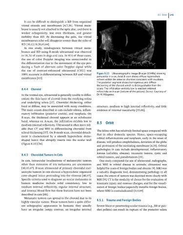
100% accurate in differentiating between RD and vitreal Figure 8.15 Ultrasonographic image (B-scan 10 MHz) showing
membranes [65]. panuveitis in a cat. Axial B-scan shows diffuse hyperechoic
echoes within the anterior chamber consistent with exudates.
The posterior segment shows homogenous and diffuse
thickening of the choroid which is distinguishable from the
8.4.4 Choroid sclera. The infiltration exhibits low to medium internal
reflectivity on A-scan (bottom of the picture). Source: Courtesy of
In the normal eye, ultrasound is generally unable to differ- Dr M. Villagrasa.
entiate the thin layer of choroid from the overlying retina
and underlying sclera [37]. Choroidal thickening, either
focal or diffuse, may be associated with many conditions. structure, medium to high internal reflectivity, and little
The main causes described in cats include edema, inflam- evidence of internal vascularity [35,68].
matory infiltration (posterior uveitis), and neoplasia. On
B‐scan, the thickened choroid appears as an echolucent
band, whereas on A‐scan, the infiltration exhibits low to 8.5 Orbit
medium internal reflectivity. Ultrasound is often more reli-
able than CT and MRI in differentiating choroidal from
scleral thickening [37]. On B‐mode scan, choroidal detach- The feline orbit has relatively limited space compared with
that in other domestic species. Hence, space‐occupying
ment is characterized by a smooth hyperechoic dome‐
shaped lesion that abruptly inserts into the ocular wall orbital inflammations and neoplasm, early in the onset of
disease, will produce exophthalmos, deviation of the globe
(Figure 8.15) [36].
and protrusion of the nictitating membrane [4,35]. Orbital
pathologies in cats include developmental, inflammatory
8.4.5 Choroidal Tumors in Cats lesions (cellulitis, abscess), traumatic lesions, cystic and
orbital tumors, and pseudotumors [35].
In cats, intraocular localizations of melanocytic tumors One study compared the use of ultrasound, radiographs,
other than extension of iris melanoma are uncommon and MRI in orbital disease in animals; ultrasound was
[40,45,67]. B‐scan ultrasound of primary choroidal mel- helpful for cases of foreign bodies and abscess [9]. MRI was
anocytic tumors in cats showed a hyperechoic organized a valuable diagnostic tool, demonstrating pathology in all
cone‐shaped lesion protruding into the vitreous [40,67]. cases; the extent of tumors was depicted more clearly with
Specific criteria used to diagnose an ocular melanoma in MRI [9]. CT is the modality of choice for the evaluation of
human medicine include solid consistency, low to traumatic injury and osseous changes, and for the visuali-
medium internal reflectivity, regular internal structure, zation of foreign bodies (especially metallic foreign bodies,
and internal blood flow but these features have not been where MRI is contraindicated) [4,9,69].
described in cats [68].
Metastatic tumors can spread to the choroid due to its
highly vascular nature. These tumors have a quite differ- 8.5.1 Trauma and Foreign Bodies
ent echographic appearance in humans; they usually Severe blunt or penetrating ocular trauma (e.g., BB or gun-
have an irregular lumpy contour, an irregular internal shot pellets) can result in rupture of the posterior sclera