
Page 144 - Feline diagnostic imaging
P. 144
8.5 rbit 143
thickening or detachment of the retina and/or choroid is are recommended to screen for metallic foreign bodies
typically present. CT has particular utility in the evaluation (Figure 8.16) [4]. Other intraocular foreign bodies, such
of orbital trauma, allowing detailed assessment of bony as plant material, produce medium amplitude echoes.
lesions with simultaneous evaluation of soft tissues, globe, CT successfully identified a linear metallic foreign body
orbit, and brain [4,71]. in one cat and a linear wood foreign body in another cat
Foreign bodies and metallic foreign bodies produce while ocular and orbital ultrasound failed to demon-
very high reflectivity on A‐scan; on B‐scan the metallic strate a plant foreign body [74–76].
foreign body produce a very echo‐dense signal that
persists at low gain settings, and there is usually marked
shadowing of the ocular and orbital structures just pos - 8.5.2 Inflammatory Processes
(Abscess, Cellulitis)
terior to the foreign body. Spherical foreign bodies (e.g.,
BB or gunshot pellets) produce a unique and specific The etiology of inflammatory/infectious diseases (orbital
echographic signal referred to as a comet tail artifact cellulitis and retrobulbar abscess) includes bacteria,
[71–73]. MRI is contraindicated in clinical cases with parasitic, fungal, pyogranulomatous, and foreign body
suspicion of metallic foreign body because the magnetic [4,74–79].
field may induce movement of the foreign body and sec- B‐mode ultrasound is helpful in the investigation of
ondary hemorrhage. In these cases, plain radiographs orbital diseases, but most authors agree that the findings
(a) (b)
*
(c)
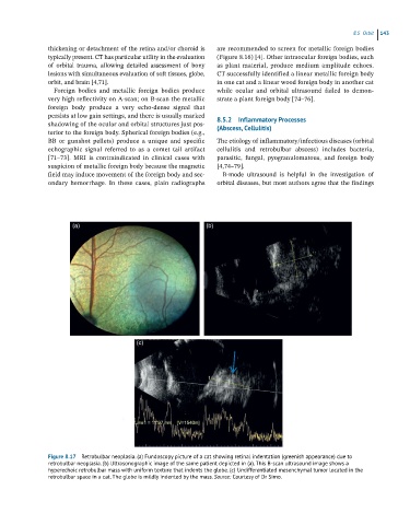
Figure 8.17 Retrobulbar neoplasia. (a) Fundoscopy picture of a cat showing retinal indentation (greenish appearance) due to
retrobulbar neoplasia. (b) Ultrasonographic image of the same patient depicted in (a). This B-scan ultrasound image shows a
hyperechoic retrobulbar mass with uniform texture that indents the globe. (c) Undifferentiated mesenchymal tumor located in the
retrobulbar space in a cat. The globe is mildly indented by the mass. Source: Courtesy of Dr Simo.