
Page 141 - Feline diagnostic imaging
P. 141
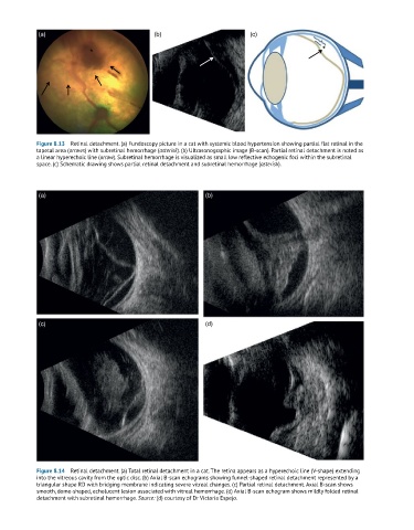
(a)                                (b)                         (c)

                            *                                                                   *


            Figure 8.13  Retinal detachment. (a) Fundoscopy picture in a cat with systemic blood hypertension showing partial flat retinal in the
            tapetal area (arrows) with subretinal hemorrhage (asterisk). (b) Ultrasonographic image (B-scan). Partial retinal detachment is noted as
            a linear hyperechoic line (arrow). Subretinal hemorrhage is visualized as small low reflective echogenic foci within the subretinal
            space. (c) Schematic drawing shows partial retinal detachment and subretinal hemorrhage (asterisk).


            (a)                                               (b)


            (c)                                               (d)


            Figure 8.14  Retinal detachment. (a) Total retinal detachment in a cat. The retina appears as a hyperechoic line (V-shape) extending
            into the vitreous cavity from the optic disc. (b) Axial B-scan echograms showing funnel-shaped retinal detachment represented by a
            triangular shape RD with bridging membrane indicating severe vitreal changes. (c) Partial retinal detachment. Axial B-scan shows
            smooth, dome-shaped, echolucent lesion associated with vitreal hemorrhage. (d) Axial B-scan echogram shows mildly folded retinal
            detachment with subretinal hemorrhage. Source: (d) courtesy of Dr Victoria Espejo.