
Page 137 - Feline diagnostic imaging
P. 137
136 8 Diseases of the Eye
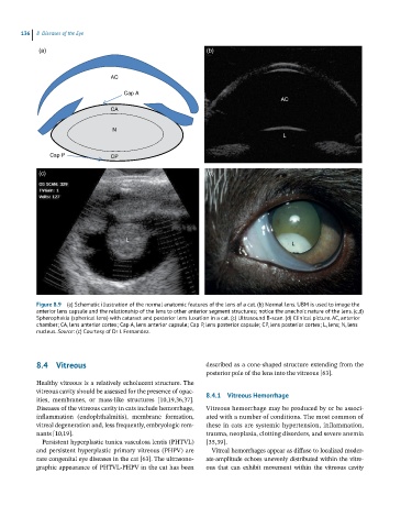
(a) (b)
AC
Cap A
AC
CA
N
L
Cap P CP
(c) (d)
L
L
Figure 8.9 (a) Schematic illustration of the normal anatomic features of the lens of a cat. (b) Normal lens. UBM is used to image the
anterior lens capsule and the relationship of the lens to other anterior segment structures; notice the anechoic nature of the lens. (c,d)
Spherophakia (spherical lens) with cataract and posterior lens luxation in a cat. (c) Ultrasound B-scan. (d) Clinical picture. AC, anterior
chamber; CA, lens anterior cortex; Cap A, lens anterior capsule; Cap P, lens posterior capsule; CP, lens posterior cortex; L, lens; N, lens
nucleus. Source: (c) Courtesy of Dr I. Fernandez.
8.4 Vitreous described as a cone‐shaped structure extending from the
posterior pole of the lens into the vitreous [63].
Healthy vitreous is a relatively echolucent structure. The
vitreous cavity should be assessed for the presence of opac- 8.4.1 Vitreous Hemorrhage
ities, membranes, or mass‐like structures [10,19,36,37].
Diseases of the vitreous cavity in cats include hemorrhage, Vitreous hemorrhage may be produced by or be associ-
inflammation (endophthalmitis), membrane formation, ated with a number of conditions. The most common of
vitreal degeneration and, less frequently, embryologic rem- these in cats are systemic hypertension, inflammation,
nants [10,19]. trauma, neoplasia, clotting disorders, and severe anemia
Persistent hyperplastic tunica vasculosa lentis (PHTVL) [35,39].
and persistent hyperplastic primary vitreous (PHPV) are Vitreal hemorrhages appear as diffuse to localized moder-
rare congenital eye diseases in the cat [63]. The ultrasono- ate‐amplitude echoes unevenly distributed within the vitre-
graphic appearance of PHTVL‐PHPV in the cat has been ous that can exhibit movement within the vitreous cavity