
Page 135 - Feline diagnostic imaging
P. 135
134 8 Diseases of the Eye
spherical protrusion), spherical lens tissue (spherophakia), Nuclear sclerosis or cataract formation increases the
or a notch‐like defect (coloboma) have all been reported internal reflectivity of the lens [3,10]. While nuclear sclero-
(Figure 8.9c,d) [58]. sis does not appear to cause detectable lesion on B‐scan,
A wide variety of pathologic conditions involving the cataracts produce increased echoes in various locations
lens can be demonstrated with ultrasound [12,18,19,44]. within an anechoic lens and internal spikes of differing
The most common acquired lens abnormalities described amplitude on A‐scan [2,4,10,12,18,36,60]. Cataracts are
in cats include cataracts, rupture of the anterior and poste- classified as incipient (<10%), immature (10 – <100%),
rior lens capsule, lens luxation, and a syndrome unique to mature (100% lens involvement), and hypermature (when
cats called aqueous misdirection glaucoma that results in reduced lens volume and wrinkled lens capsule are visual-
anterior displacement of the lens [50,59]. ized) [12]. Lens thickness may be increased in a mature
cataract, while hypermature cataracts are often thin on
ultrasound, because the lens protein has liquefied and
reabsorbed [3,10,12]. According to their location within
the lens, assessed with ultrasound, cataracts will be catego-
rized as capsular, cortical (anterior, posterior), nuclear, or
complete (cortico‐nuclear cataract) (Figure 8.10) [12].
The lens anterior or posterior capsular rupture may be
associated with anterior uveitis, osmotic cataract, trauma,
and synechia [12]. Irregularity of the lens capsule and the
suspected presence of cortical lens material on the outer
AC L V
surface of the capsule have been described as a typical
ultrasonographic appearance of lens capsule rupture
using high‐resolution ultrasound 20 MHz (Figure 8.11)
[12,20,44,61].
Lens position can easily be assessed by ultrasound as it lies
evenly between the ciliary bodies [4]. Lens luxation has been
described with microphthalmia and spherophakia in cats
or can be associated with trauma, chronic uveitis, neoplasia,
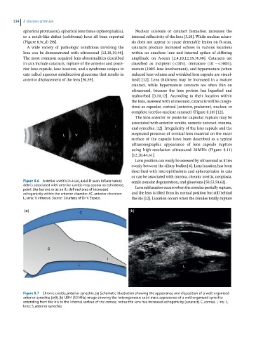
Figure 8.6 Anterior uveitis in a cat, axial B-scan. Inflammatory senile zonular degeneration, and glaucoma [50,53,54,62].
debris associated with anterior uveitis may appear as echodense, Lens subluxation occurs when the zonules partially rupture,
point-like lesions or as an ill-defined area of increased
echogenicity within the anterior chamber. AC, anterior chamber; and the lens is tilted from its normal position but still behind
L, lens; V, vitreous. Source: Courtesy of Dr V. Espejo. the iris [12]. Luxation occurs when the zonules totally rupture
(a) C (b)
C
S
S I
I
L
L
Figure 8.7 Chronic uveitis, anterior synechia. (a) Schematic illustration showing the appearance and disposition of a well-organized
anterior synechia (left). (b) UBM (50 MHz) image showing the heterogeneous solid mass appearance of a well-organized synechia
extending from the iris to the internal surface of the cornea; notice the lens has increased echogenicity (cataract). C, cornea; I, iris; L,
lens; S, anterior synechia.