
Page 132 - Feline diagnostic imaging
P. 132
8.2 Iffaaaatooy aIn eoofastiic Diseases ffeictiIng the Iteoioo engaeIt 131
(a) (b)
C A P R S C A P R
S
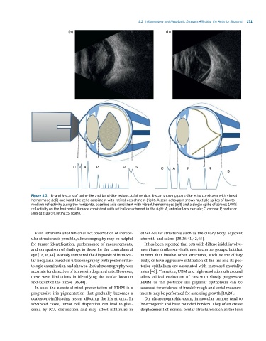
Figure 8.2 B- and A-scans of point-like and band-like lesions. Axial vertical B-scan showing point-like echo consistent with vitreal
hemorrhage (left) and band-like echo consistent with retinal detachment (oinght). A-scan echogram shows multiple spikes of low to
medium reflectivity along the horizontal baseline axis consistent with vitreal hemorrhages (left) and a single spike of almost 100%
reflectivity on the horizontal A-mode consistent with retinal detachment in the right. A, anterior lens capsule; C, cornea; P, posterior
lens capsule; R, retina; S, sclera.
Even for animals for which direct observation of intraoc- other ocular structures such as the ciliary body, adjacent
ular structures is possible, ultrasonography may be helpful choroid, and sclera [35,36,41,42,45].
for tumor identification, performance of measurements, It has been reported that cats with diffuse iridal involve-
and comparison of findings to those for the contralateral ment have similar survival times to control groups, but that
eye [18,36,44]. A study compared the diagnosis of intraocu- tumors that involve other structures, such as the ciliary
lar neoplasia based on ultrasonography with posterior his- body, or have aggressive infiltration of the iris and its pos-
tologic examination and showed that ultrasonography was terior epithelium are associated with increased mortality
accurate for detection of tumors in dogs and cats. However, rates [46]. Therefore, UBM and high‐resolution ultrasound
there were limitations in identifying the ocular location allow critical evaluation of cats with slowly progressive
and extent of the tumor [36,44]. FDIM as the posterior iris pigment epithelium can be
In cats, the classic clinical presentation of FDIM is a assessed for evidence of breakthrough and serial measure-
progressive iris pigmentation that gradually becomes a ments may be performed for assessing growth [18,20].
coalescent‐infiltrating lesion affecting the iris stroma. In On ultrasonographic exam, intraocular tumors tend to
advanced cases, tumor cell dispersion can lead to glau- be echogenic and have rounded borders. They often create
coma by ICA obstruction and may affect infiltrates in displacement of normal ocular structures such as the lens