
Page 134 - Feline diagnostic imaging
P. 134
8.3 ens 133
C
AC
L
I
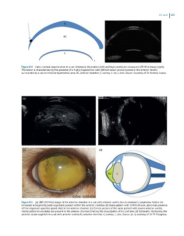
Figure 8.4 Calcic corneal degeneration in a cat. Schematic illustration (left) and high-resolution ultrasound (35 MHz) image (oinght).
The lesion is characterized by the presence of a highly hyperechoic well-defined lesion (arrow) located in the anterior stroma
surrounded by a low to medium hyperechoic area. AC, anterior chamber; C, cornea; I, iris; L, lens. Source: Courtesy of Dr Victoria Espejo.
(a) C (b)
AC
AC L
L
(c) (d)
AC L
Figure 8.5 (a) UBM (50 MHz) image of the anterior chamber in a cat with anterior uveitis due to metastatic lymphoma. Notice the
increased echogenicity (well organized) present within the anterior chamber. (b) Same patient with 10 MHz B-scan, abnormal presence
of fine dispersed opacities (point-like) in the anterior chamber. (c) Clinical picture of the same patient with severe anterior uveitis,
visible yellowish exudates are present in the anterior chamber, limiting the visualization of iris and lens. (d) Schematic illustrating the
anterior ocular segment in a cat with anterior uveitis.AC, anterior chamber; C, cornea; L, lens. Source: (a–c) courtesy of Dr M. Villagrasa.