
Page 138 - Feline diagnostic imaging
P. 138
8.4 Vitreous 137
(a) (b)
L
AC
V L
(c) (d)
L
AC V
L
V
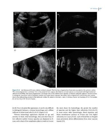
Figure 8.10 (a) Ultrasound B-scan, anterior cortical cataract. The lens has a hyperechoic focal area localized in the anterior cortex.
The strong hyperechoic curvilinear line is the posterior lens capsule. (b) Anterior cortical cataract as visualized with high-resolution
ultrasound (35 MHz); the strong hyperechoic curvilinear line is the anterior lens capsule. (c) Mature complete cataract. The entire lens
is echogenic, consistent with a complete cataract. (d) Hypermature cataract; the entire lens is hyperechoic and thinner than normal
due to the reabsorbing process in the lenticular cortex. AC, anterior chamber; L, lens; V, vitreous. Source: (a) Courtesy of Dr I. Fernandez.
(b–d) Courtesy of Dr Victoria Espejo.
[4,38]. Due to its point‐like appearance, it can be very difficult the more dense the hemorrhage, the greater the number
to distinguish between a vitreous hemorrhage and a diffuse of opacities and the higher their reflectivity [10,19,36,37].
infiltrate of white blood cells [12,19,35] (Figure 8.12). In more severe older hemorrhages, blood organizes and
Vitreous hemorrhage appearance depends on age and forms membranous surfaces on B‐scan and even higher
severity. In fresh, mild hemorrhage, dots and short lines of reflectivity on A‐scan [37,64]. Lack of blood flow on Doppler
low reflective mobile vitreous opacities are displayed on B‐ exam potentially allows differentiation from other vascular
scan, and a chain of low‐amplitude spikes is noted on A‐scan; masses [19].