
Page 143 - Feline diagnostic imaging
P. 143
142 8 Diseases of the Eye
(a) (b)
*
(c) (d)
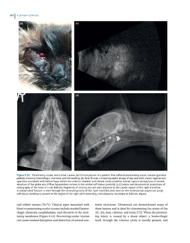
Figure 8.16 Penetrating ocular and orbital trauma. (a) Clinical picture of a patient that suffered penetrating ocular trauma (gunshot
pellets) showing hemorrhagic chemosis and lid swelling. (b) Axial B-scan ultrasonographic image of eye and orbit shows hyperechoic
opacities consistent with hemorrhage within the anterior chamber and vitreal cavity posterior scleral rupture (arrow), loss of normal
structure of the globe and diffuse hyperechoic echoes in the orbital soft tissue (asterisk). (c,d) Lateral and dorsoventral projections of
radiography of the head of a cat. Ballistic fragments of varying size are seen adjacent to the caudal aspect of the right mandible.
A comminuted fracture is seen through the coronoid process of the right mandible, best seen on the ventrodorsal projection. Large
soft tissue swelling is present in the region of the right orbit extending craniolaterally secondary to ballistic impact.
and orbital trauma [70,71]. Clinical signs associated with tomic structures. Ultrasound can demonstrated many of
blunt or penetrating ocular trauma include marked hemor- these lesions and is ideal for determining the status of the
rhagic chemosis, exophthalmia, and elevation of the nicti- AC, iris, lens, vitreous, and retina [71]. When the penetrat-
tating membrane (Figure 8.16). Penetrating ocular injuries ing injury is caused by a sharp object, a hemorrhagic
can cause marked disruption and distortion of normal ana- track through the vitreous cavity is usually present, and