
Page 185 - Feline diagnostic imaging
P. 185
11.3 Computed CoCogrmphy Cofuptf telit pCgra 187
(b)
(a)
(c) (d)
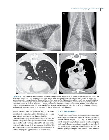
Figure 11.14 (a,b) Lateral (a) and ventrodorsal (b) thoracic images of a cat presented for cough, weight loss, and lethargy. A large soft
tissue mass is identified in the right caudal lung lobe. Several small pulmonary nodules are barely visible. (c) Transverse CT image
(postcontrast, using a lung window) of the caudal thorax of the same cat. The right caudal lung lobe mass (mass) is noted just lateral
to the caudal vena cava (vc), closely associated with the right diaphragmatic crus. In addition, several soft tissue pulmonary nodules
are present (arrows). A, aorta. (d) Transverse CT image (postcontrast, using a soft tissue window) through the hilus of the lung of the
same cat. The tracheobronchial lymph nodes are markedly enlarged (LN). E, esophagus; T, trachea. At necropsy, a primary pulmonary
carcinoma, with metastasis to the lungs and tracheobronchial lymph nodes, was diagnosed.
viscous effusions such as pyothorax may be unilateral. 11.3.7 Pneumothorax
Exudate in the pleural space (pyothorax) may also be locu-
lated rather than symmetric and dependent [4]. Free air in the pleural space creates a nonattenuating space
between parietal and visceral pleural layers in the nonde-
Computed tomography lymphangiography has been per-
formed using ultrasound‐guided injection of mesenteric pendent thorax (Figure 11.18) [20]. CT may be used to
search for pulmonary bullae in cases of spontaneous pneu-
lymph nodes with contrast, followed by CT scans of the
contrast‐filled cisterna chyli and thoracic duct [34]. This mothorax. Bullae are visualized as round, thin‐walled cavi-
ties with low pulmonary parenchymal density. In dogs, the
procedure could be done in cases of chylothorax to evalu-
ate the integrity and appearance of the thoracic duct. sensitivity and positive predictive value of CT for detection