
Page 180 - Feline diagnostic imaging
P. 180
182 11 Advanced Imaging Modalities
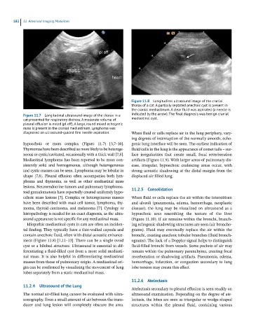
Figure 11.8 Longitudinal ultrasound image of the cranial
thorax of a cat. A partially septated anechoic cyst is present in
the cranial mediastinum. A clear fluid was aspirated (a needle is
Figure 11.7 Longitudinal ultrasound image of the thorax in a indicated by the arrow). The final diagnosis was benign cranial
cat presented for respiratory distress. A moderate volume of mediastinal cyst.
pleural effusion is noted (pl eff). A large, round mixed echogenic
mass is present in the cranial mediastinum. Lymphoma was
diagnosed on ultrasound-guided fine needle aspiration. When fluid or cells replace air in the lung periphery, vary-
ing degrees of interruption of the normally smooth, echo-
hypoechoic or more complex (Figure 11.7) [3,7–10]. genic lung interface will be seen. The earliest indication of
Thymomas have been described as more likely to be heteroge- fluid/cells in the lung is the appearance of comet tails – sur-
neous or cystic/cavitated, occasionally with a thick wall [7,8]. face irregularities that create small, focal reverberation
Mediastinal lymphoma has been reported to be more con- artifacts (Figure 11.9). With larger areas of pulmonary dis-
sistently solid and homogeneous, although heterogeneous ease, irregular, hypoechoic coalescing areas occur, with
and cystic masses can be seen. Lymphoma may be lobular in strong acoustic shadowing at the distal margin from the
shape [7,8]. Pleural effusion often accompanies both lym- displaced air‐filled lung.
phoma and thymoma, as well as other mediastinal mass
lesions. Neuroendocrine tumors and pulmonary lymphoma-
toid granulomatosis have reportedly created uniformly hypo- 11.2.5 Consolidation
echoic mass lesions [7]. Complex or heterogeneous masses When fluid or cells replace the air within the interstitium
have been described with mast cell tumor, lymphoma, thy- and alveoli (pneumonia, edema, hemorrhage, neoplastic
moma, thyroid carcinoma, and melanoma [7]. Cytology or disease), the lung may be visualized on ultrasound as a
histopathology is needed for an exact diagnosis, as the ultra- hypoechoic area resembling the texture of the liver
sound appearance is not specific for any mediastinal mass. (Figure 11.10). If air remains within the bronchi, branch-
Idiopathic mediastinal cysts in cats are often an inciden- ing echogenic shadowing structures are seen (air broncho-
tal finding. They typically have a thin‐walled capsule and grams). Fluid may eventually replace the air within the
contain anechoic fluid, often with distal acoustic enhance- bronchi, creating anechoic tubular branches (fluid bronch-
ment (Figure 11.8) [7,11–13]. There can be a single ovoid ograms). The lack of a Doppler signal helps to distinguish
cyst or a bilobed structure. Ultrasound is essential in dif- fluid‐filled bronchi from vessels. Some pockets of air may
ferentiating a fluid‐filled cyst from a more solid mediasti- remain within the pulmonary parenchyma, creating focal
nal mass. It is also helpful in differentiating mediastinal reverberation or shadowing artifacts. Pneumonia, edema,
masses from those of pulmonary origin. A mediastinal ori- hemorrhage, infarction, or congestion secondary to lung
gin can be confirmed by visualizing the movement of lung lobe torsion may create this effect.
lobes separately from a static mediastinal mass.
11.2.6 Atelectasis
11.2.4 Ultrasound of the Lung
Atelectasis secondary to pleural effusion is seen readily on
The normal air‐filled lung cannot be evaluated with ultra- ultrasound examination. Depending on the degree of ate-
sonography. Even a small amount of air between the trans- lectasis, the lobes are seen as triangular or wedge‐shaped
ducer and lung lesion will completely obscure the area. structures within the pleural fluid, containing various