
Page 175 - Feline diagnostic imaging
P. 175
176 10 Normal Cardiovascular Imaging
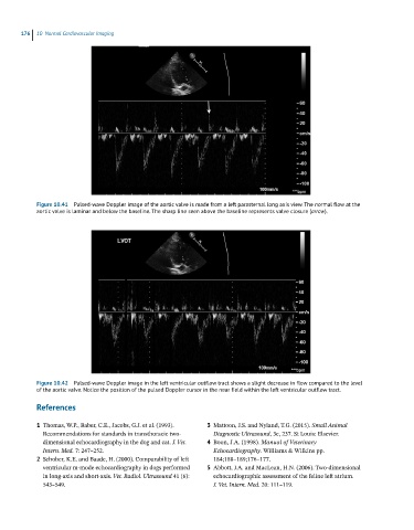
Figure 10.41 Pulsed-wave Doppler image of the aortic valve is made from a left parasternal long axis view. The normal flow at the
aortic valve is laminar and below the baseline. The sharp line seen above the baseline represents valve closure (arroe).
Figure 10.42 Pulsed-wave Doppler image in the left ventricular outflow tract shows a slight decrease in flow compared to the level
of the aortic valve. Notice the position of the pulsed Doppler cursor in the near field within the left ventricular outflow tract.
References
1 Thomas, W.P., Baber, C.E., Jacobs, G.J. et al. (1993). 3 Mattoon, J.S. and Nyland, T.G. (2015). Small Animal
Recommendations for standards in transthoracic two‐ Diagnostic Ultrasound, 3e, 237. St Louis: Elsevier.
dimensional echocardiography in the dog and cat. J. Vet. 4 Boon, J.A. (1998). Manual of Veterinary
Intern. Med. 7: 247–252. Echocardiography. Williams & Wilkins pp.
2 Schober, K.E. and Baade, H. (2000). Comparability of left 184;188–189;176–177.
ventricular m‐mode echocardiography in dogs performed 5 Abbott, J.A. and MacLean, H.N. (2006). Two‐dimensional
in long‐axis and short‐axis. Vet. Radiol. Ultrasound 41 (6): echocardiographic assessment of the feline left atrium.
543–549. J. Vet. Intern. Med. 20: 111–119.