
Page 170 - Feline diagnostic imaging
P. 170
10.7 Doppler 171
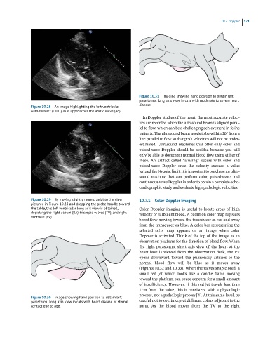
Figure 10.31 Imaging showing hand position to obtain left
parasternal long axis view in cats with moderate to severe heart
Figure 10.28 An image highlighting the left ventricular disease.
outflow tract (LVOT) as it approaches the aortic valve (Ao).
In Doppler studies of the heart, the most accurate veloci-
ties are recorded when the ultrasound beam is aligned paral-
lel to flow, which can be a challenging achievement in feline
patients. The ultrasound beam needs to be within 20° from a
line parallel to flow so that peak velocities will not be under-
estimated. Ultrasound machines that offer only color and
pulsed‐wave Doppler should be avoided because you will
only be able to document normal blood flow using either of
these. An artifact called “aliasing” occurs with color and
pulsed‐wave Doppler once the velocity exceeds a value
termed the Nyquist limit. It is important to purchase an ultra-
sound machine that can perform color, pulsed‐wave, and
continuous‐wave Doppler in order to obtain a complete echo-
cardiographic study and evaluate high pathologic velocities.
Figure 10.29 By moving slightly more cranial to the view 10.7.1 Color Doppler Imaging
pictured in Figure 10.23 and dropping the probe handle toward
the table, this left ventricular long axis view is obtained, Color Doppler imaging is useful to locate areas of high
depicting the right atrium (RA), tricuspid valves (TV), and right velocity or turbulent blood. A common color map registers
ventricle (RV).
blood flow moving toward the transducer as red and away
from the transducer as blue. A color bar representing the
selected color map appears on an image when color
Doppler is activated. Think of the top of the image as an
observation platform for the direction of blood flow. When
the right parasternal short axis view of the heart at the
heart base is viewed from the observation deck, the PV
opens downward toward the pulmonary arteries so the
normal blood flow will be blue as it moves away
(Figures 10.32 and 10.33). When the valves snap closed, a
small red jet which looks like a candle flame moving
toward the platform can cause concern for a small amount
of insufficiency. However, if this red jet travels less than
1 cm from the valve, this is consistent with a physiologic
process, not a pathologic process [4]. At this same level, be
Figure 10.30 Image showing hand position to obtain left
parasternal long axis view in cats with heart disease or sternal careful not to overinterpret different colors adjacent to the
contact due to age. aorta. As the blood moves from the TV to the right