
Page 169 - Feline diagnostic imaging
P. 169
170 10 Normal Cardiovascular Imaging
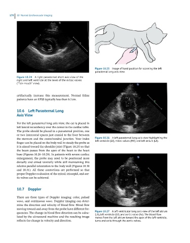
Figure 10.25 Image of hand position for scanning the left
parasternal long axis view.
Figure 10.24 A right parasternal short axis view of the
right and left ventricle at the level of the mitral valves
(“fish-mouth” view).
artifactually increase this measurement. Normal feline
patients have an EPSS typically less than 0.2 cm.
10.6 Left Parasternal Long
Axis View
For the left parasternal long axis view, the cat is placed in
left lateral recumbency over the cutout in the cardiac table.
The probe should be placed in a parasternal position, one
or two intercostal spaces just cranial to the liver between
the sternum and the costochondral junction. Your index Figure 10.26 A left parasternal long axis view highlighting the
finger can be placed on the body wall to steady the probe as left ventricle (LV), mitral valves (MV), and left atrium (LA).
it is aimed toward the shoulder joint (Figure 10.25) so that
the beam passes from the apex of the heart to the heart
base (Figures 10.26–10.29). In patients with severe cardiac
enlargement, the probe may need to be positioned more
dorsally and aimed ventrally while still maintaining this
relative parallel orientation to the body wall (Figures 10.30
and 10.31). All these contortions are performed so that
proper Doppler evaluation of the mitral, tricuspid, and aor-
tic valves can be achieved.
10.7 Doppler
There are three types of Doppler imaging: color, pulsed
wave, and continuous wave. Doppler imaging can deter-
mine the direction and velocity of blood flow. Blood flow
moving toward and away from the probe have different fre-
quencies. The change in blood flow direction can be calcu- Figure 10.27 A left ventricular long axis view of the left atrium
(LA), left ventricle (LV), and aortic valve (Ao). The blood flow
lated by the ultrasound machine and the resulting image moves from the left atrium toward the apex of the left ventricle,
reflects the change in velocity and direction. turns and exits through the aortic valves.