
Page 174 - Feline diagnostic imaging
P. 174
Acknoeledgment 175
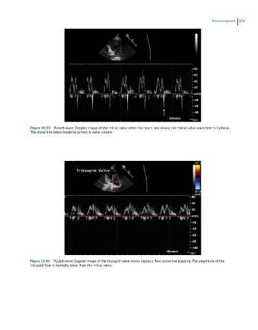
Figure 10.39 Pulsed-wave Doppler image of the mitral valve when the heart rate slows; the mitral valve waveform is biphasic.
The sharp line below baseline (arroe) is valve closure.
Figure 10.40 Pulsed-wave Doppler image of the tricuspid valve shows biphasic flow above the baseline. The amplitude of the
tricuspid flow is normally lower than the mitral valve.