
Page 178 - Feline diagnostic imaging
P. 178
180 11 Advanced Imaging Modalities
separate structure only when fluid is present on both sides 11.2.1 Pleural Effusion
(pleural and peritoneal effusion). The diaphragm should be Pleural effusion, if sufficient in volume, creates an excel-
carefully and completely evaluated when diaphragmatic
hernia is suspected, using both subcostal and intercostal lent acoustic window, allowing evaluation of the mediasti-
num, chest wall, and lungs. Cranially, bilateral pleural
windows. A mirror image artifact is usually present in nor-
mal cats, creating the appearance of liver on both sides of effusion outlines the midline cranial mediastinal tissue,
while more caudally, pleural fluid will enhance visualiza-
the diaphragm.
tion of the heart and caudal vena cava (Figure 11.3).
Depending on the cellular content, the fluid appears ane-
choic or slightly echogenic, displacing the lung lobes away
from the chest wall and diaphragm.
Occasionally, pleural effusion is discovered incidentally
while performing abdominal ultrasonography (Figure 11.3).
Fluid can be seen cranial to the diaphragm while scanning
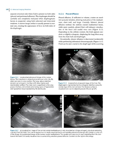
Figure 11.1 Longitudinal ultrasound image of the normal
thorax. The transducer is aligned perpendicular to the ribs (R),
which are seen in cross-section. The lung–pleura interface
(arrow) is an echogenic interface with deep shadow and Figure 11.3 Longitudinal ultrasound image of the liver. The
reverberation artifact. The chest wall is superficial to the lung– diaphragm is represented by the echogenic interface along the
pleura interface, and contains alternating layers of hyperechoic cranial edge of the liver (arrows). The thoracic surface is
and hypoechoic tissue representing fat and muscle. visualized well due to adjacent pleural effusion (pl eff).
(a) (b)
Figure 11.2 (a) Longitudinal image of the normal cranial mediastinum (cr ms), visualized as a linear, echogenic structure extending
cranially from the heart. The cranial mediastinum is easily visualized due to surrounding pleural effusion (pl eff). Cranial is to the left
of the image. (b) Longitudinal image of the normal caudal mediastinum. The caudal vena cava (cvc) is seen extending from the liver
toward the heart. It is easily visualized due to surrounding anechoic pleural effusion. Cranial is to the right in this image.