
Page 181 - Feline diagnostic imaging
P. 181
11.2 ormal Anatomy 183
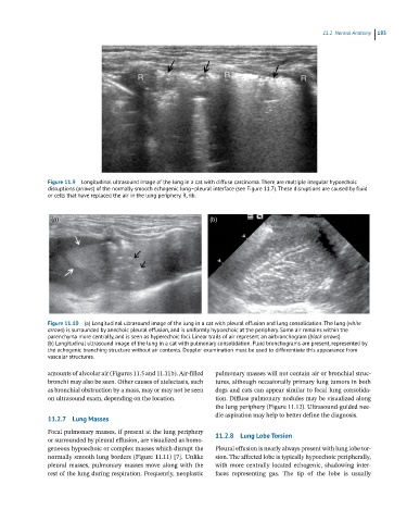
Figure 11.9 Longitudinal ultrasound image of the lung in a cat with diffuse carcinoma. There are multiple irregular hypoechoic
disruptions (arrows) of the normally smooth echogenic lung–pleural interface (see Figure 11.7). These disruptions are caused by fluid
or cells that have replaced the air in the lung periphery. R, rib.
(a) (b)
Figure 11.10 (a) Longitudinal ultrasound image of the lung in a cat with pleural effusion and lung consolidation. The lung (white
arrows) is surrounded by anechoic pleural effusion, and is uniformly hypoechoic at the periphery. Some air remains within the
parenchyma more centrally, and is seen as hyperechoic foci. Linear trails of air represent an airbronchogram (black arrows).
(b) Longitudinal ultrasound image of the lung in a cat with pulmonary consolidation. Fluid bronchograms are present, represented by
the echogenic branching structure without air contents. Doppler examination must be used to differentiate this appearance from
vascular structures.
amounts of alveolar air (Figures 11.5 and 11.11b). Air‐filled pulmonary masses will not contain air or bronchial struc-
bronchi may also be seen. Other causes of atelectasis, such tures, although occasionally primary lung tumors in both
as bronchial obstruction by a mass, may or may not be seen dogs and cats can appear similar to focal lung consolida-
on ultrasound exam, depending on the location. tion. Diffuse pulmonary nodules may be visualized along
the lung periphery (Figure 11.12). Ultrasound‐guided nee-
dle aspiration may help to better define the diagnosis.
11.2.7 Lung Masses
Focal pulmonary masses, if present at the lung periphery 11.2.8 Lung Lobe Torsion
or surrounded by pleural effusion, are visualized as homo-
geneous hypoechoic or complex masses which disrupt the Pleural effusion is nearly always present with lung lobe tor-
normally smooth lung borders (Figure 11.11) [7]. Unlike sion. The affected lobe is typically hypoechoic peripherally,
pleural masses, pulmonary masses move along with the with more centrally located echogenic, shadowing inter-
rest of the lung during respiration. Frequently, neoplastic faces representing gas. The tip of the lobe is usually