
Page 186 - Feline diagnostic imaging
P. 186
188 11 Advanced Imaging Modalities
(a) (b)
(c)
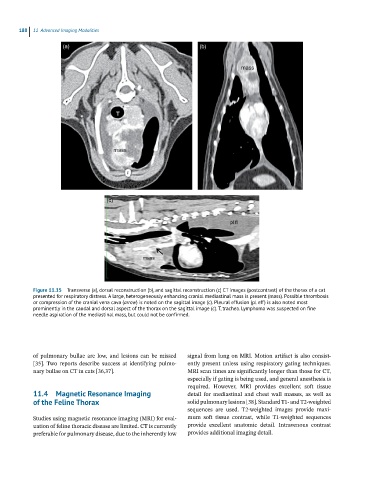
Figure 11.15 Transverse (a), dorsal reconstruction (b), and sagittal reconstruction (c) CT images (postcontrast) of the thorax of a cat
presented for respiratory distress. A large, heterogeneously enhancing cranial mediastinal mass is present (mass). Possible thrombosis
or compression of the cranial vena cava (arrow) is noted on the sagittal image (c). Pleural effusion (pl eff) is also noted most
prominently in the caudal and dorsal aspect of the thorax on the sagittal image (c). T, trachea. Lymphoma was suspected on fine
needle aspiration of the mediastinal mass, but could not be confirmed.
of pulmonary bullae are low, and lesions can be missed signal from lung on MRI. Motion artifact is also consist-
[35]. Two reports describe success at identifying pulmo- ently present unless using respiratory gating techniques.
nary bullae on CT in cats [36,37]. MRI scan times are significantly longer than those for CT,
especially if gating is being used, and general anesthesia is
required. However, MRI provides excellent soft tissue
11.4 Magnetic Resonance Imaging detail for mediastinal and chest wall masses, as well as
of the Feline Thorax solid pulmonary lesions [38]. Standard T1‐ and T2‐weighted
sequences are used. T2‐weighted images provide maxi-
Studies using magnetic resonance imaging (MRI) for eval- mum soft tissue contrast, while T1‐weighted sequences
uation of feline thoracic disease are limited. CT is currently provide excellent anatomic detail. Intravenous contrast
preferable for pulmonary disease, due to the inherently low provides additional imaging detail.