
Page 191 - Feline diagnostic imaging
P. 191
(a) (b)
(c) (d)
(e)
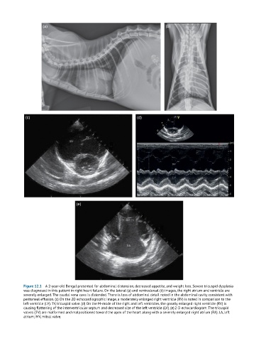
Figure 12.1 A 2-year-old Bengal presented for abdominal distension, decreased appetite, and weight loss. Severe tricuspid dysplasia
was diagnosed in this patient in right heart failure. On the lateral (a) and ventrodorsal (b) images, the right atrium and ventricle are
severely enlarged. The caudal vena cava is distended. There is loss of abdominal detail noted in the abdominal cavity consistent with
peritoneal effusion. (c) On the 2D echocardiographic image, a moderately enlarged right ventricle (RV) is noted in comparison to the
left ventricle (LV). TV, tricuspid valve. (d) On the M-mode of the right and left ventricles, the greatly enlarged right ventricle (RV) is
causing flattening of the interventricular septum and decreased size of the left ventricle (LV). (e) 2-D echocardiogram. The tricuspid
valves (TV) are malformed and malpositioned toward the apex of the heart along with a severely enlarged right atrium (RA). LA, left
atrium; MV, mitral valve.